Copyright
©The Author(s) 2024.
World J Gastrointest Oncol. Apr 15, 2024; 16(4): 1256-1267
Published online Apr 15, 2024. doi: 10.4251/wjgo.v16.i4.1256
Published online Apr 15, 2024. doi: 10.4251/wjgo.v16.i4.1256
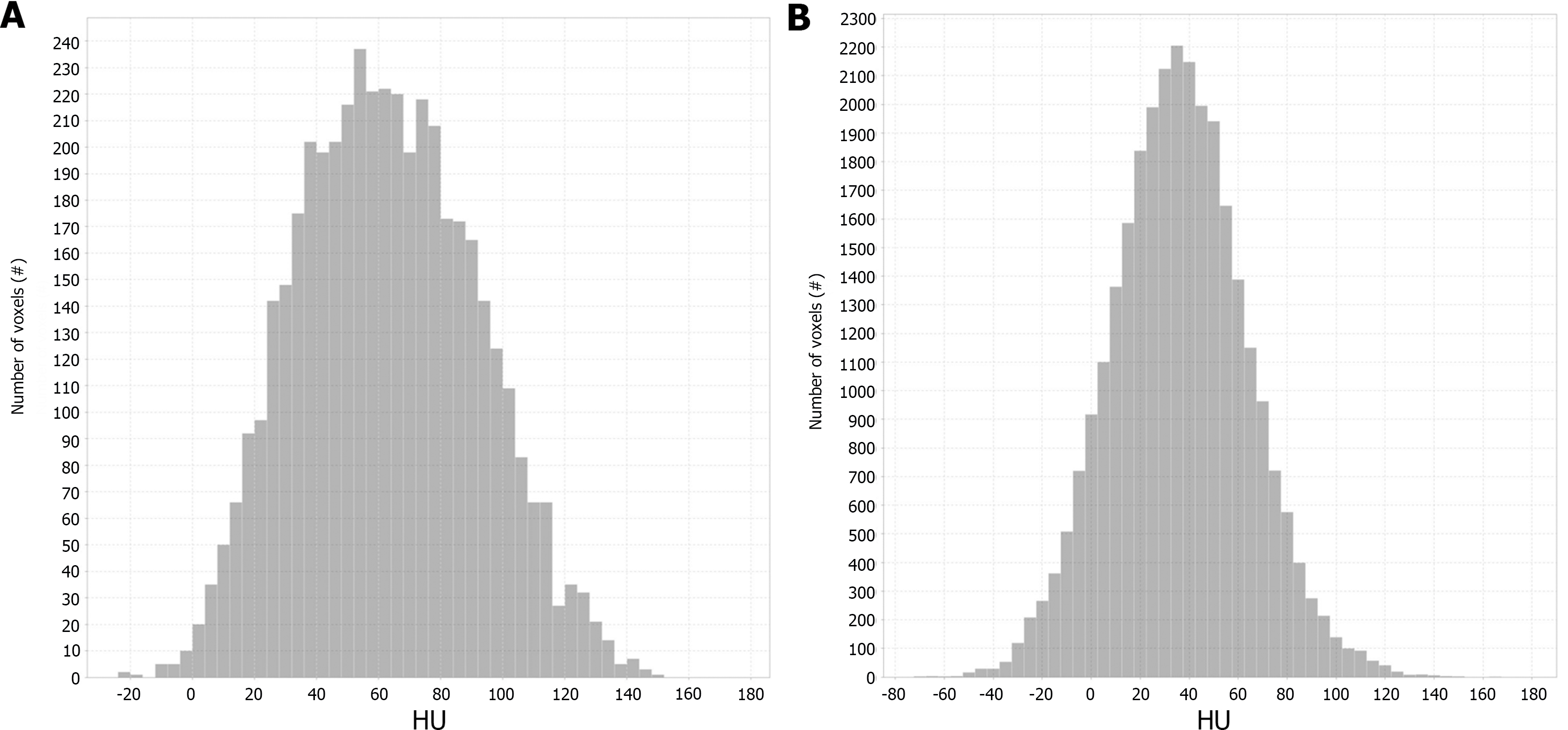
Figure 3 Histograms of two representative cases of pancreatic ductal adenocarcinoma.
A and B: Stage IB pancreatic ductal adenocarcinoma (PDAC), A vs stage IV PDAC, B, which showed a marked difference. The X-axis indicates the gray level (HU). The Y-axis indicates the number of voxels.
- Citation: Ren S, Qian LC, Cao YY, Daniels MJ, Song LN, Tian Y, Wang ZQ. Computed tomography-based radiomics diagnostic approach for differential diagnosis between early- and late-stage pancreatic ductal adenocarcinoma. World J Gastrointest Oncol 2024; 16(4): 1256-1267
- URL: https://www.wjgnet.com/1948-5204/full/v16/i4/1256.htm
- DOI: https://dx.doi.org/10.4251/wjgo.v16.i4.1256


