Copyright
©The Author(s) 2024.
World J Gastrointest Oncol. Apr 15, 2024; 16(4): 1256-1267
Published online Apr 15, 2024. doi: 10.4251/wjgo.v16.i4.1256
Published online Apr 15, 2024. doi: 10.4251/wjgo.v16.i4.1256
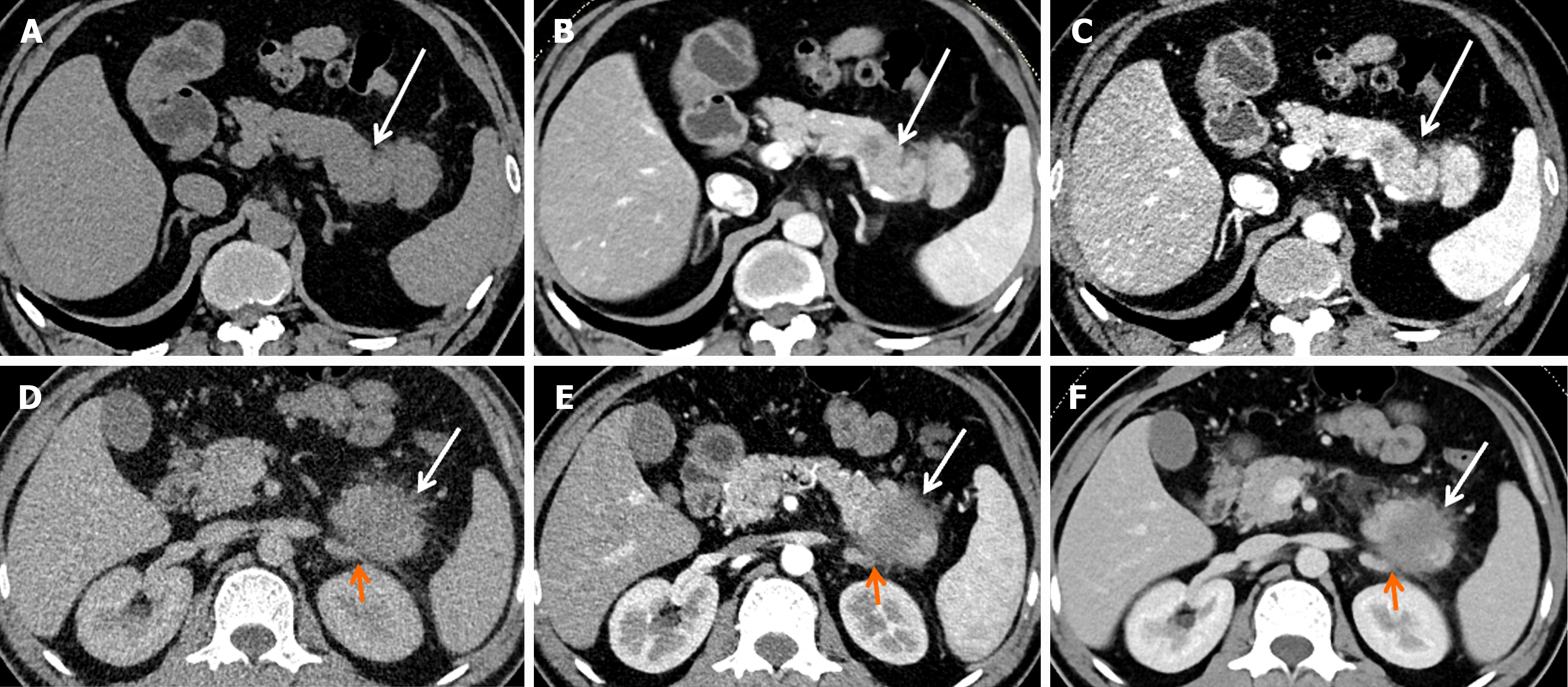
Figure 2 Computed tomography images of two representative cases of pancreatic ductal adenocarcinoma.
A-C: A 54-year-old man with stage IB pancreatic ductal adenocarcinoma (PDAC). Computed tomography (CT) imaging showed a well-defined, predominantly solid tumor (white arrow), 3.0 cm in diameter in the tail of the pancreas. No obvious lymph node metastasis, vascular invasion, or distal metastasis was found. TNM classification: T2N0M0 (IB); D-F: A 44-year-old man with stage IV PDAC. CT imaging showed an ill-defined, predominantly solid tumor (white arrow), 4.1 cm in diameter in the tail of the pancreas. The left adrenal gland (orange arrow) was grossly invaded by the tumor. TNM classification: T3N1M1 (IV).
- Citation: Ren S, Qian LC, Cao YY, Daniels MJ, Song LN, Tian Y, Wang ZQ. Computed tomography-based radiomics diagnostic approach for differential diagnosis between early- and late-stage pancreatic ductal adenocarcinoma. World J Gastrointest Oncol 2024; 16(4): 1256-1267
- URL: https://www.wjgnet.com/1948-5204/full/v16/i4/1256.htm
- DOI: https://dx.doi.org/10.4251/wjgo.v16.i4.1256


