Copyright
©The Author(s) 2024.
World J Gastrointest Oncol. Mar 15, 2024; 16(3): 819-832
Published online Mar 15, 2024. doi: 10.4251/wjgo.v16.i3.819
Published online Mar 15, 2024. doi: 10.4251/wjgo.v16.i3.819
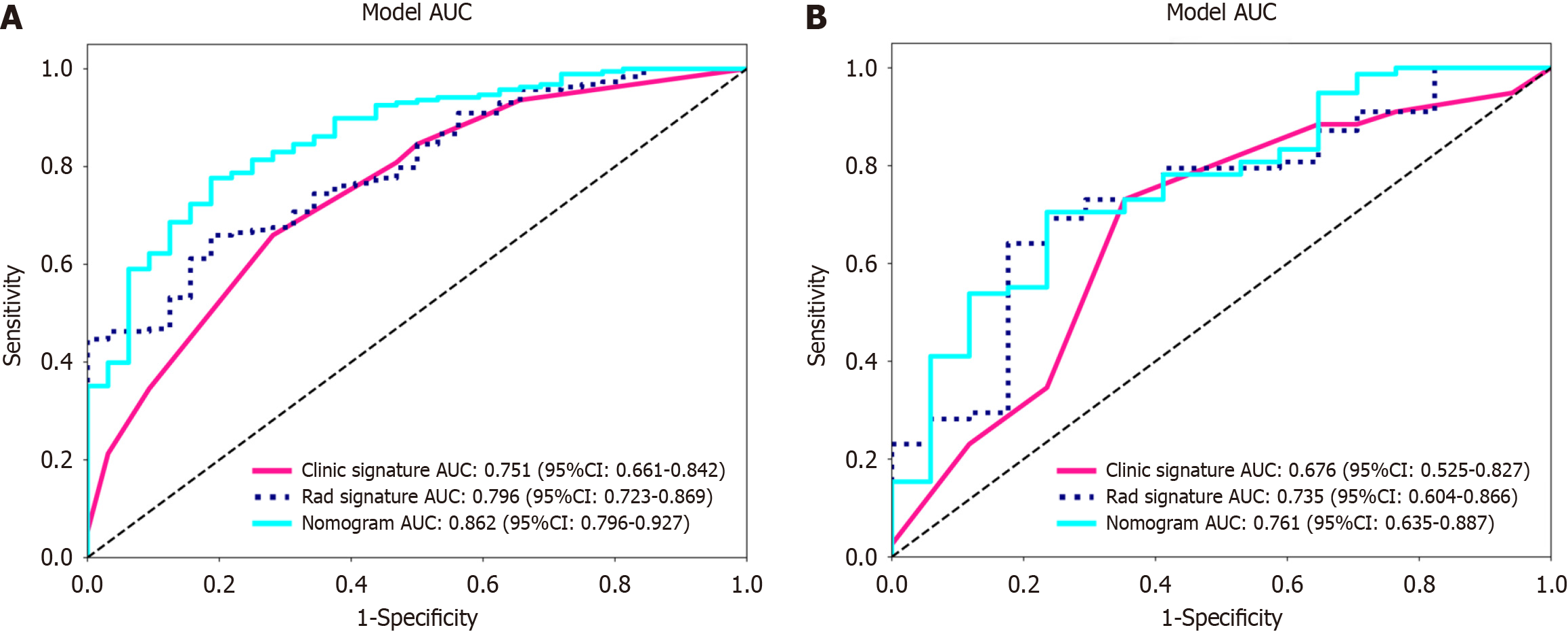
Figure 8 Receiver operating characteristic curves of the radiomic model, clinical model and radiomic-clinical model.
A: The area under the curve (AUC) of the three models (clinical, radiomic, and radiomic-clinical model) in the training cohort were 0.751 (95%CI: 0.661-0.842), 0.796 (95%CI: 0.723-0.869), and 0.862 (95%CI: 0.796-0.927), respectively. B: The AUC of the three models (clinical, radiological, and radiomic-clinical model) in the validation cohort were 0.676 (95%CI: 0.525-0.827), 0.735 (95%CI: 0.604-0.866), and 0.761 (95%CI: 0.635-0.887), respectively. ROC: Receiver operating characteristic; AUC: Area under the curve.
- Citation: Zheng HD, Huang QY, Huang QM, Ke XT, Ye K, Lin S, Xu JH. T2-weighted imaging-based radiomic-clinical machine learning model for predicting the differentiation of colorectal adenocarcinoma. World J Gastrointest Oncol 2024; 16(3): 819-832
- URL: https://www.wjgnet.com/1948-5204/full/v16/i3/819.htm
- DOI: https://dx.doi.org/10.4251/wjgo.v16.i3.819


