Copyright
©The Author(s) 2024.
World J Gastrointest Oncol. Mar 15, 2024; 16(3): 773-786
Published online Mar 15, 2024. doi: 10.4251/wjgo.v16.i3.773
Published online Mar 15, 2024. doi: 10.4251/wjgo.v16.i3.773
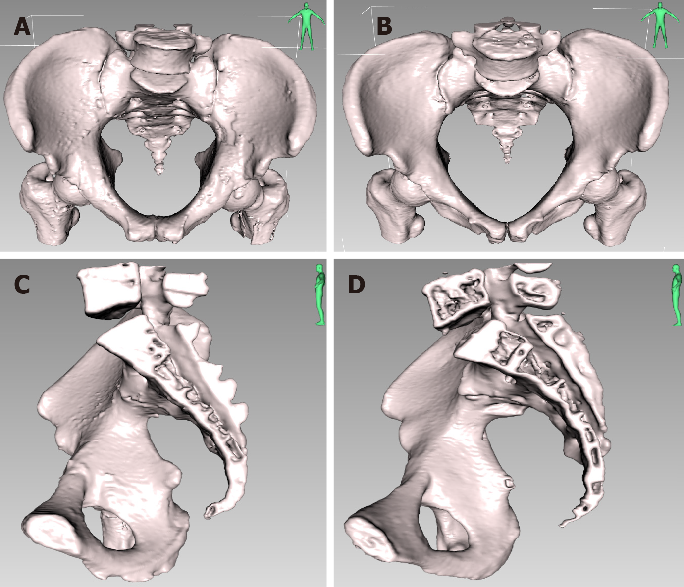
Figure 4 Front and lateral view of the three-dimensional pelvic reconstruction between male and female patients in the forward tilt position and the mid-sagittal position respectively.
A: Front view of the three-dimensional (3D) pelvic reconstruction in a male patient in the forward tilt position; B: Front view of the 3D pelvic reconstruction in a female patient in the forward tilt position; C: Lateral view of the 3D pelvic reconstruction in a male patient in the mid-sagittal position; D: Lateral view of the 3D pelvic reconstruction in a female patient in the mid-sagittal position. The male pelvis is deep and narrow, with a forward tilt, straighter sacrum, and a higher overall curvature. The female pelvis is wide and shallow, with a backward tilt and a smaller overall curvature.
- Citation: Zhou XC, Ke FY, Dhamija G, Chen H, Wang Q. Study on sex differences and potential clinical value of three-dimensional computerized tomography pelvimetry in rectal cancer patients. World J Gastrointest Oncol 2024; 16(3): 773-786
- URL: https://www.wjgnet.com/1948-5204/full/v16/i3/773.htm
- DOI: https://dx.doi.org/10.4251/wjgo.v16.i3.773


