Copyright
©The Author(s) 2024.
World J Gastrointest Oncol. Mar 15, 2024; 16(3): 699-715
Published online Mar 15, 2024. doi: 10.4251/wjgo.v16.i3.699
Published online Mar 15, 2024. doi: 10.4251/wjgo.v16.i3.699
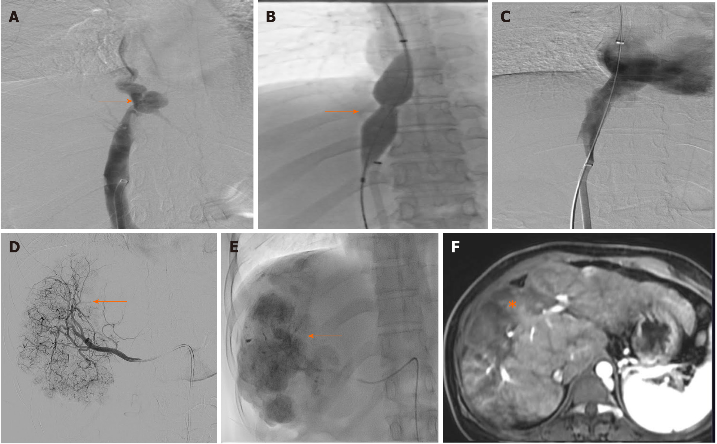
Figure 4 Digital subtraction spot images.
A-F: Digital subtraction spot images showed short segment narrowing of inferior vena cava (A, arrow) which was dilated using 20 mm × 40 mm balloon catheter (B, arrow), post angioplasty angiogram (C) good flow across the inferior vena cava without any residual narrowing. Selective right hepatic angiogram showed tumor blush (D, arrow), which was treated using lipiodol transarterial chemoembolization (E, arrow), follow-up magnetic resonance imaging after transarterial chemoembolization showed no residual enhancing lesion in the treated lesion (F, asterisk).
- Citation: Agarwal A, Biswas S, Swaroop S, Aggarwal A, Agarwal A, Jain G, Elhence A, Vaidya A, Gupte A, Mohanka R, Kumar R, Mishra AK, Gamanagatti S, Paul SB, Acharya SK, Shukla A, Shalimar. Clinical profile and outcomes of hepatocellular carcinoma in primary Budd-Chiari syndrome. World J Gastrointest Oncol 2024; 16(3): 699-715
- URL: https://www.wjgnet.com/1948-5204/full/v16/i3/699.htm
- DOI: https://dx.doi.org/10.4251/wjgo.v16.i3.699


