Copyright
©The Author(s) 2024.
World J Gastrointest Oncol. Mar 15, 2024; 16(3): 670-686
Published online Mar 15, 2024. doi: 10.4251/wjgo.v16.i3.670
Published online Mar 15, 2024. doi: 10.4251/wjgo.v16.i3.670
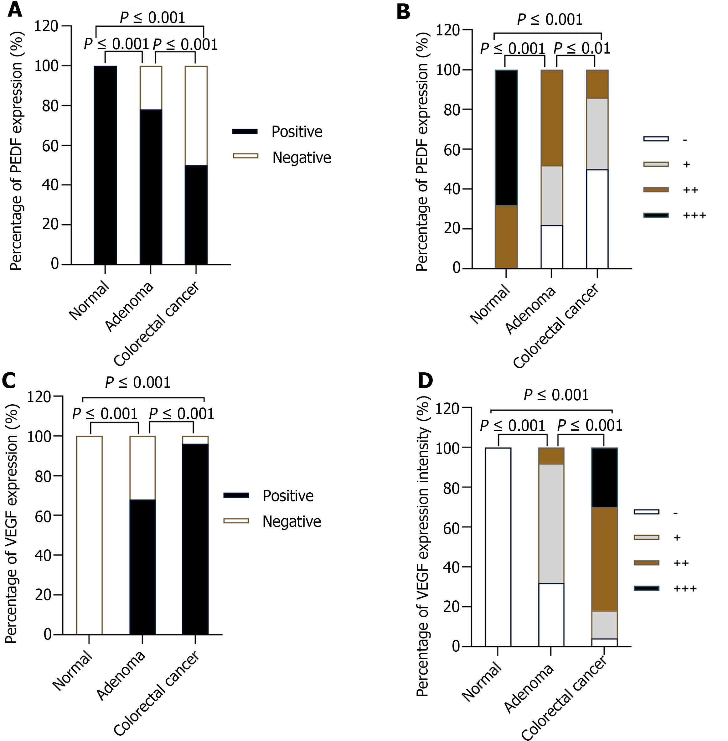
Figure 2 Comparison of positive expression rate and expression intensity of pigment epithelium-derived factors and vascular endothelial growth factors in normal control group, adenoma group and colorectal cancer group.
A: Positive expression rate of pigment epithelium-derived factors (PEDF) in the three groups; B: Expression intensity of PEDF in the three groups; C: Positive expression rate of vascular endothelial growth factors (VEGF) in the three groups; D: Expression intensity of VEGF in the three groups. n = 50 (normal control group), n = 50 (adenoma group), n = 50 (colorectal cancer group). PEDF: Pigment epithelium-derived factors; VEGF: Vascular endothelial growth factors.
- Citation: Yang Y, Wen W, Chen FL, Zhang YJ, Liu XC, Yang XY, Hu SS, Jiang Y, Yuan J. Expression and significance of pigment epithelium-derived factor and vascular endothelial growth factor in colorectal adenoma and cancer. World J Gastrointest Oncol 2024; 16(3): 670-686
- URL: https://www.wjgnet.com/1948-5204/full/v16/i3/670.htm
- DOI: https://dx.doi.org/10.4251/wjgo.v16.i3.670


