Copyright
©The Author(s) 2024.
World J Gastrointest Oncol. Mar 15, 2024; 16(3): 614-629
Published online Mar 15, 2024. doi: 10.4251/wjgo.v16.i3.614
Published online Mar 15, 2024. doi: 10.4251/wjgo.v16.i3.614
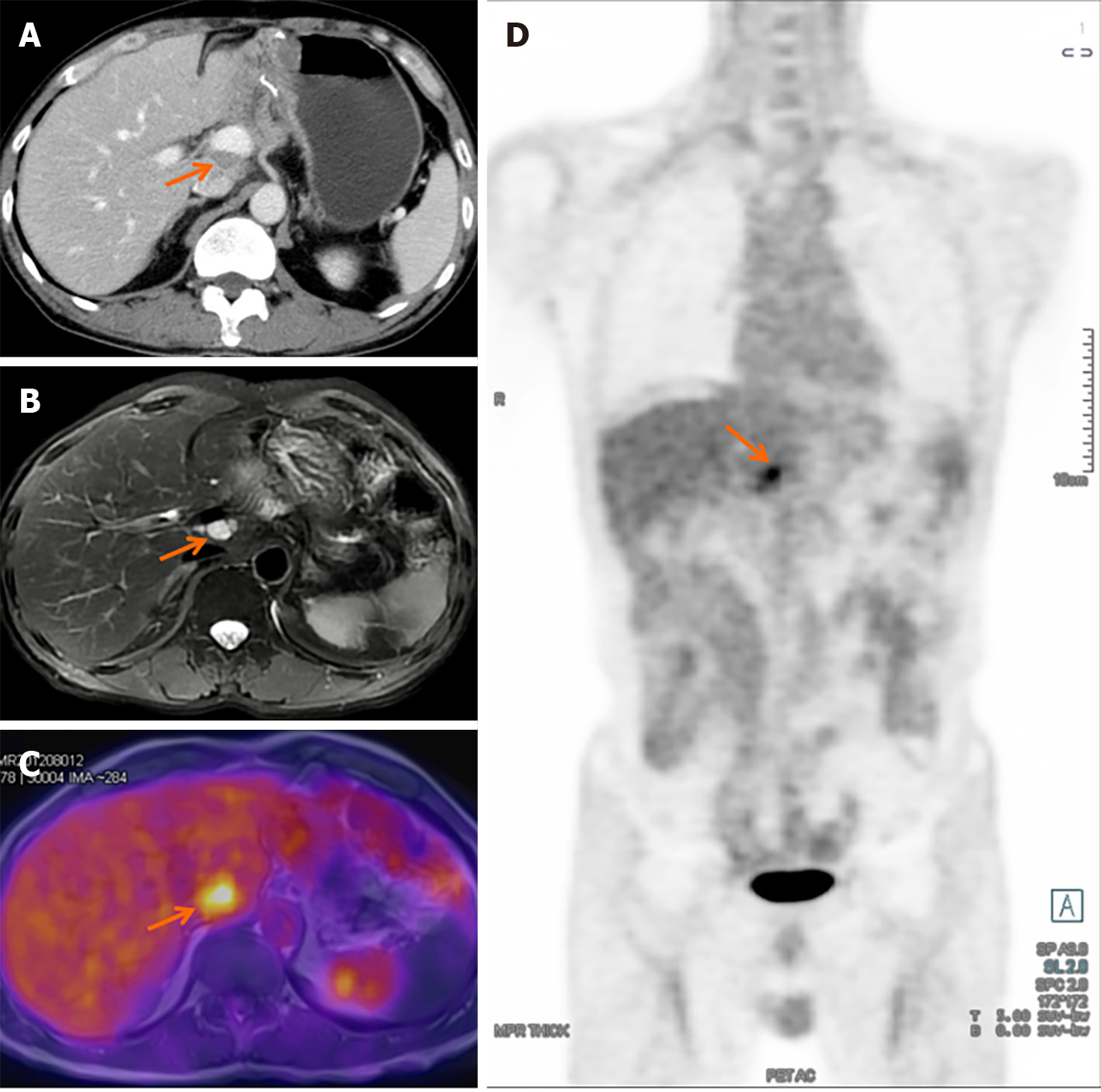
Figure 6 Detection of recurrence in solid pseudopapillary tumor of the pancreas.
A: Abdominal enhanced computed tomography scan revealed a hypodense mass between the portal vein and inferior vena cava; B: T2WI magnetic resonance imaging (MRI) demonstrated a high signal mass located at the hepatic hilum; C and D: Axial-fused (C) and coronal-fused (D) positron emission tomography/MRI showed that the lesion had increased fluorodeoxyglucose uptake.
- Citation: Xu YC, Fu DL, Yang F. Unraveling the enigma: A comprehensive review of solid pseudopapillary tumor of the pancreas. World J Gastrointest Oncol 2024; 16(3): 614-629
- URL: https://www.wjgnet.com/1948-5204/full/v16/i3/614.htm
- DOI: https://dx.doi.org/10.4251/wjgo.v16.i3.614


