Copyright
©The Author(s) 2024.
World J Gastrointest Oncol. Feb 15, 2024; 16(2): 543-549
Published online Feb 15, 2024. doi: 10.4251/wjgo.v16.i2.543
Published online Feb 15, 2024. doi: 10.4251/wjgo.v16.i2.543
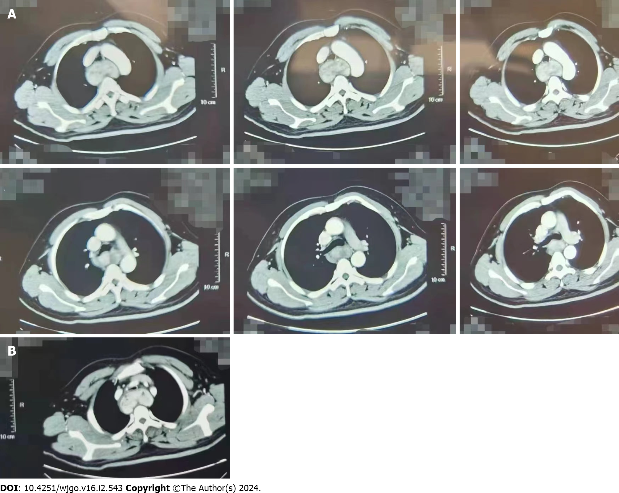
Figure 1 Computed tomography of the neck.
A: Posterior superior mediastinal esophageal travel area rich in blood supply occupying lesions, consider extraesophageal and intertracheal tumor lesions; B: This image shows the most obvious level of tracheal compression.
- Citation: Yu JJ, Pei HS, Meng Y. Large isolated fibrous tumors in the upper esophagus: A case report. World J Gastrointest Oncol 2024; 16(2): 543-549
- URL: https://www.wjgnet.com/1948-5204/full/v16/i2/543.htm
- DOI: https://dx.doi.org/10.4251/wjgo.v16.i2.543


