Copyright
©The Author(s) 2024.
World J Gastrointest Oncol. Dec 15, 2024; 16(12): 4746-4752
Published online Dec 15, 2024. doi: 10.4251/wjgo.v16.i12.4746
Published online Dec 15, 2024. doi: 10.4251/wjgo.v16.i12.4746
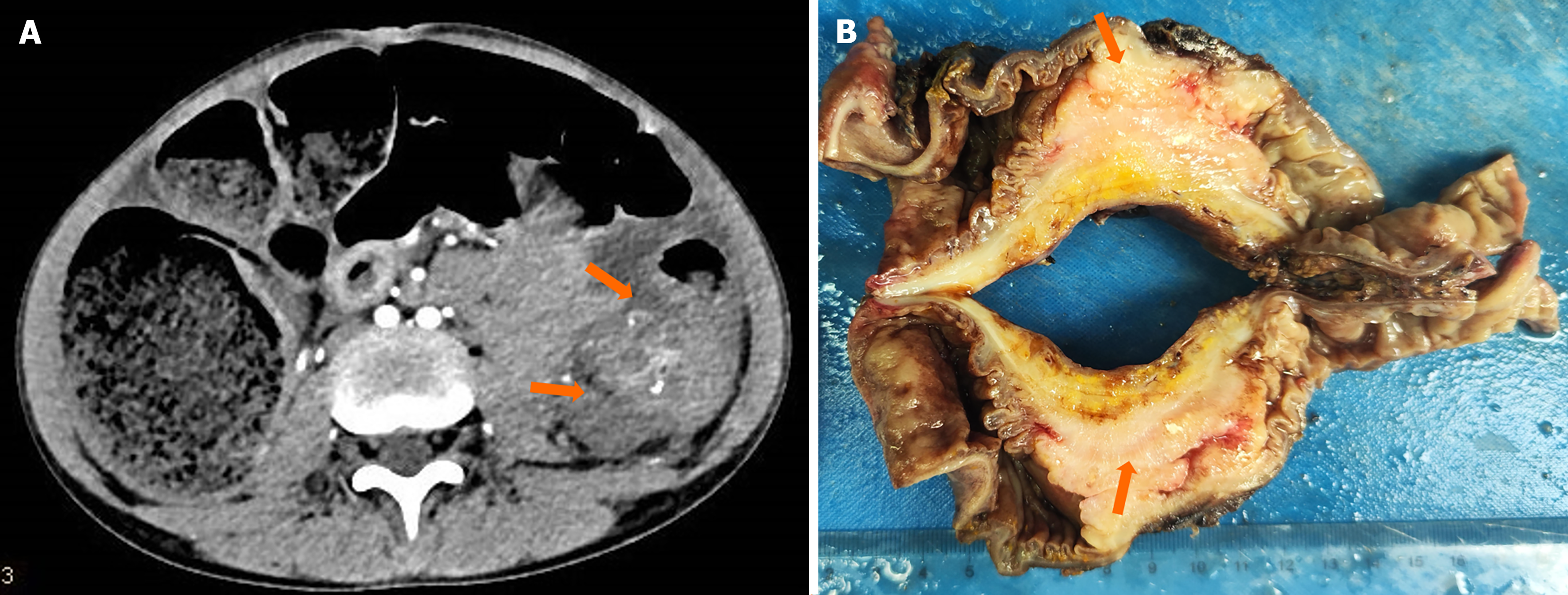
Figure 1 Tumor imaging data and surgical gross specimens.
A: Computed tomography scan revealed irregular masses of soft tissue density in the transverse colon, exhibiting heterogeneous density and multiple calcifications. The lesion exerted pressure on the adjacent descending colon, resulting in obstruction of the proximal transverse colon (orange arrowhead); B: Gross specimen of the tumor (orange arrowhead).
- Citation: Lv L, Song YH, Gao Y, Pu SQ, A ZX, Wu HF, Zhou J, Xie YC. Signet-ring cell carcinoma of the transverse colon in a 10-year-old girl: A case report. World J Gastrointest Oncol 2024; 16(12): 4746-4752
- URL: https://www.wjgnet.com/1948-5204/full/v16/i12/4746.htm
- DOI: https://dx.doi.org/10.4251/wjgo.v16.i12.4746


