Copyright
©The Author(s) 2024.
World J Gastrointest Oncol. Oct 15, 2024; 16(10): 4264-4273
Published online Oct 15, 2024. doi: 10.4251/wjgo.v16.i10.4264
Published online Oct 15, 2024. doi: 10.4251/wjgo.v16.i10.4264
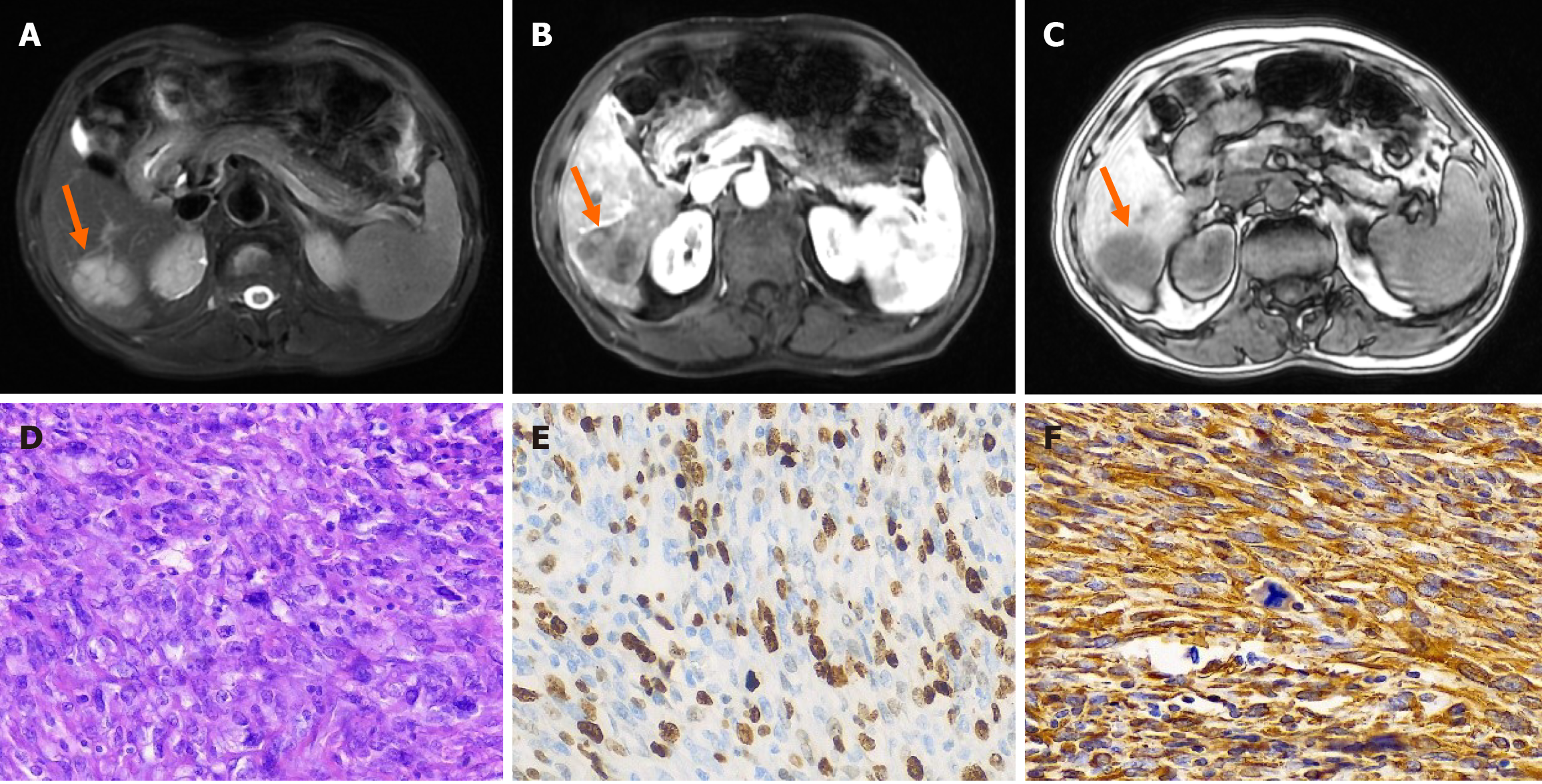
Figure 1 Magnetic resonance imaging and pathological results of primary hepatic fibrosarcoma.
A: Magnetic resonance imaging (MRI) T2 weighted imaging of the compressed fat sequence revealed high signal density (arrow); B: MRI revealed mild heterogeneity with modestly distinct margins during the arterial phase (arrow); C: The lesion in the hepatobiliary phase showed low signal intensity (arrow); D: Hematoxylin and eosin staining 400 ×; E: Ki-67 positivity rate of approximately 60%, immunohistochemical staining 400 ×; F: Vimin (+), immunohistochemical staining 400 ×.
- Citation: Zhao Y, Bie YK, Zhang GY, Feng YB, Wang F. Rare and lacking typical clinical symptoms of liver tumors: Four case reports. World J Gastrointest Oncol 2024; 16(10): 4264-4273
- URL: https://www.wjgnet.com/1948-5204/full/v16/i10/4264.htm
- DOI: https://dx.doi.org/10.4251/wjgo.v16.i10.4264


