Copyright
©The Author(s) 2024.
World J Gastrointest Oncol. Oct 15, 2024; 16(10): 4138-4145
Published online Oct 15, 2024. doi: 10.4251/wjgo.v16.i10.4138
Published online Oct 15, 2024. doi: 10.4251/wjgo.v16.i10.4138
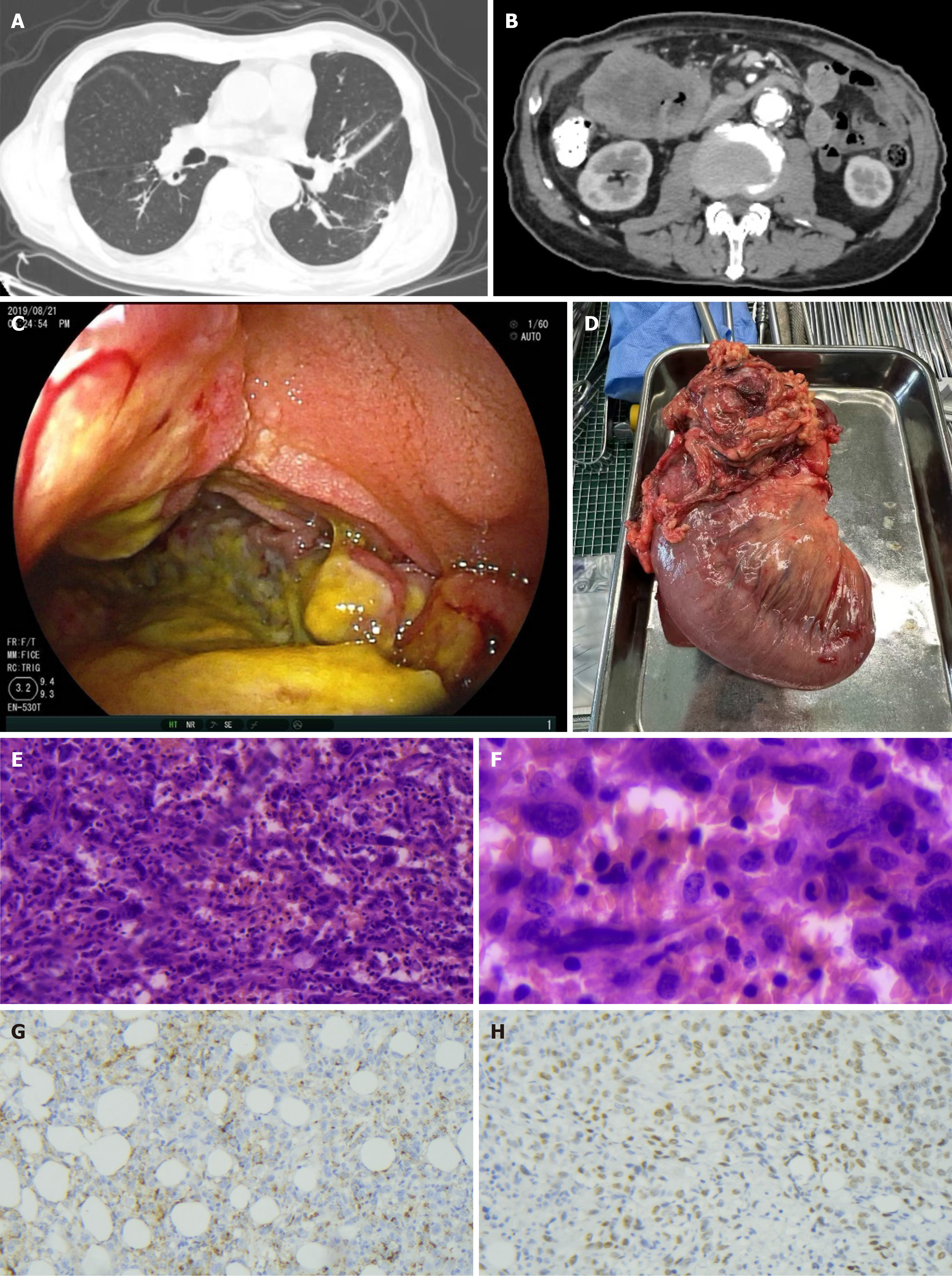
Figure 1 Computed tomography, surgical specimens and pathological images of small intestinal metastases from lung cancer.
A: The lung computed tomography examination indicate postoperative changes in lung cancer; B: The abdominal computed tomography scan indicate a huge small intestine mass in the upper right abdomen; C: Ulcerative metastases are visible on small intestinal endoscopy; D: Intussusception and obstruction resulting from small intestinal metastases are detected by surgical specimen; E: Hematoxylin and eosin staining of small intestine metastatic tumor tissue (× 100); F: Hematoxylin and eosin staining of small intestine metastatic tumor tissue (× 400); G: Immunohistochemical staining of small intestine metastatic tumor tissue shows positive expression of Napsin A (× 100); H: Immunohistochemical staining of small intestine metastatic tumor tissue shows positive expression of thyroid transcription factor 1 (× 100).
- Citation: Zhang Z, Liu J, Yu PF, Yang HR, Li JY, Dong ZW, Shi W, Gu GL. Clinicopathological analysis of small intestinal metastasis from extra-abdominal/extra-pelvic malignancy. World J Gastrointest Oncol 2024; 16(10): 4138-4145
- URL: https://www.wjgnet.com/1948-5204/full/v16/i10/4138.htm
- DOI: https://dx.doi.org/10.4251/wjgo.v16.i10.4138


