Copyright
©The Author(s) 2023.
World J Gastrointest Oncol. Jun 15, 2023; 15(6): 925-942
Published online Jun 15, 2023. doi: 10.4251/wjgo.v15.i6.925
Published online Jun 15, 2023. doi: 10.4251/wjgo.v15.i6.925
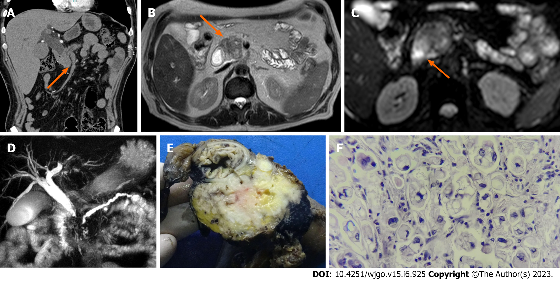
Figure 1 A 62-year-old male patient with a history of abdominal pain and jaundice.
A: Contrast-enhanced abdominal tomography shows a poorly enhanced hypocaptured lesion (orange arrow); B: Magnetic resonance imaging (MRI) in the T2 sequence shows a hypointense lesion (orange arrow); C: MRI in the diffusion sequence shows a lesion with restriction (orange arrow); D: In magnetic resonance cholangiopancreatography, the double duct sign is evident; E: Macroscopic specimen of the head of the pancreas in which there is a whitish mass; F: Slides report poorly differentiated pancreatic ductal adenocarcinoma of the pancreas with high-grade signet ring cells with perineural infiltration and invasion of the duodenum and ampulla of Vater.
- Citation: Tornel-Avelar AI, Velarde Ruiz-Velasco JA, Pelaez-Luna M. Pancreatic cancer, autoimmune or chronic pancreatitis, beyond tissue diagnosis: Collateral imaging and clinical characteristics may differentiate them. World J Gastrointest Oncol 2023; 15(6): 925-942
- URL: https://www.wjgnet.com/1948-5204/full/v15/i6/925.htm
- DOI: https://dx.doi.org/10.4251/wjgo.v15.i6.925


