Copyright
©The Author(s) 2023.
World J Gastrointest Oncol. Nov 15, 2023; 15(11): 2041-2048
Published online Nov 15, 2023. doi: 10.4251/wjgo.v15.i11.2041
Published online Nov 15, 2023. doi: 10.4251/wjgo.v15.i11.2041
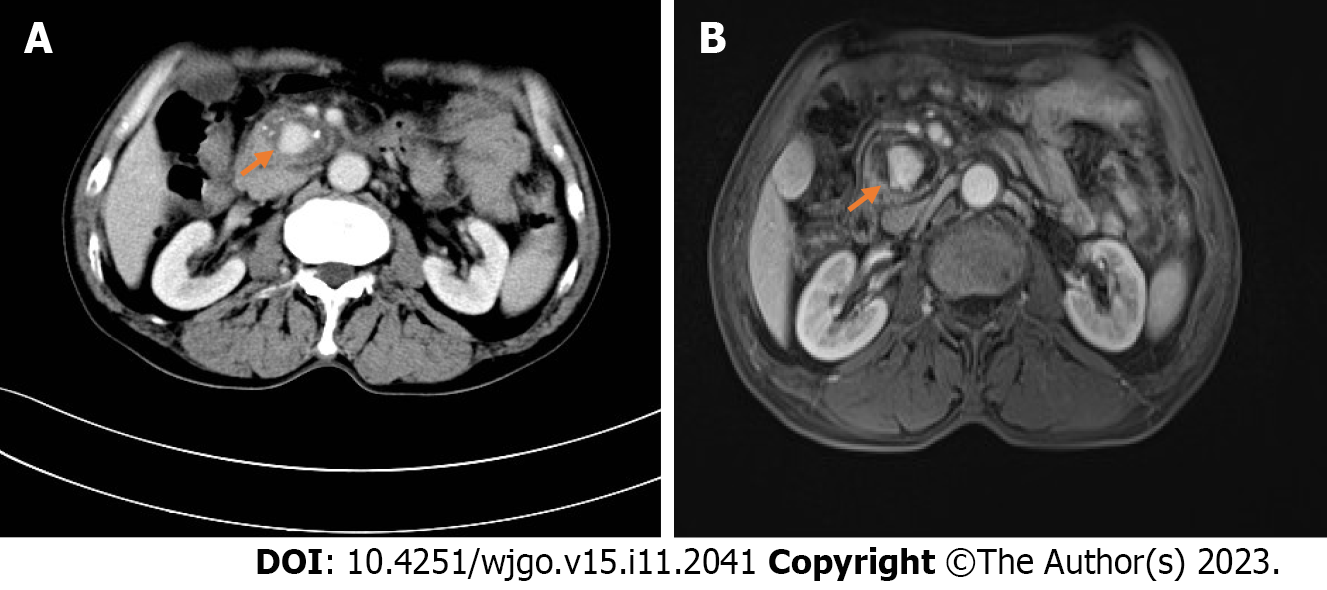
Figure 4 Computed tomography and magnetic resonance imaging venous phase imaging.
A: Axial view of contrast-enhanced computed tomography reveal delayed enhancement in the venous phase (orange arrow); B: Axial view of contrast-enhanced magnetic resonance imaging show delayed enhancement in the venous phase (orange arrow).
- Citation: Yang Y, Liu XM, Li HP, Xie R, Tuo BG, Wu HC. Pancreatic pseudoaneurysm mimicking pancreatic tumor: A case report and review of literature. World J Gastrointest Oncol 2023; 15(11): 2041-2048
- URL: https://www.wjgnet.com/1948-5204/full/v15/i11/2041.htm
- DOI: https://dx.doi.org/10.4251/wjgo.v15.i11.2041


