Copyright
©The Author(s) 2023.
World J Gastrointest Oncol. Jan 15, 2023; 15(1): 205-214
Published online Jan 15, 2023. doi: 10.4251/wjgo.v15.i1.205
Published online Jan 15, 2023. doi: 10.4251/wjgo.v15.i1.205
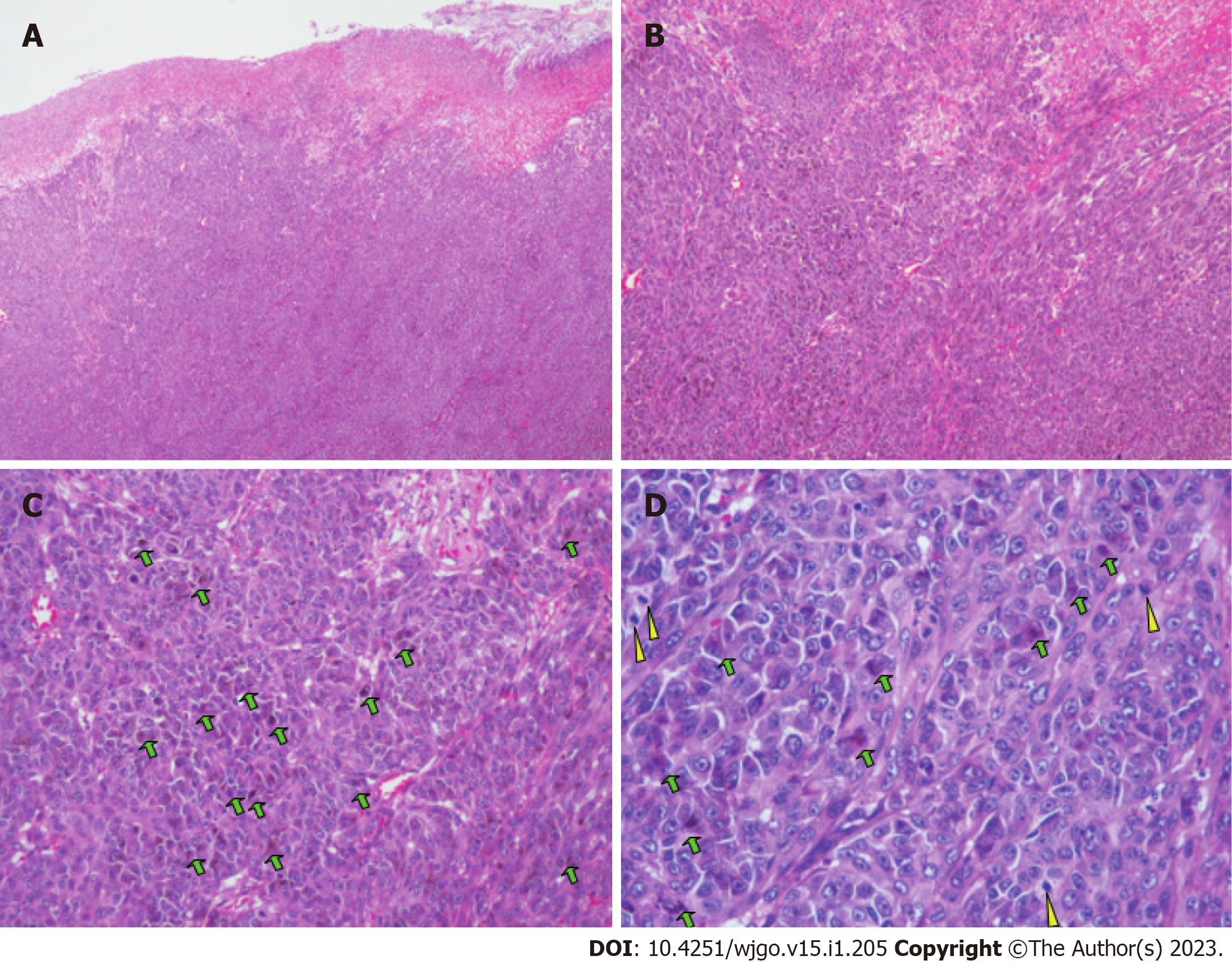
Figure 7 Histopathological findings of the resected small intestine after the second surgery.
A: Representative specimen showing that diffuse growth of tumor tissue affected the subserous layer. Hematoxylin and eosin, × 4; B: Tumor cells presented with a patchy distribution. Hematoxylin and eosin, × 10; C: Tumors had large nuclei, prominent nucleoli, and red cytoplasm. Some tumor cells showed pigment granules in the cytoplasm (green arrows). Hematoxylin and eosin, × 20; D: Some tumor cells showed pigment granules in the cytoplasm (green arrows). Epithelioid tumor cells contained mitotic figures (yellow arrows). Hematoxylin and eosin, × 40.
- Citation: Fan WJ, Cheng HH, Wei W. Surgical treatments of recurrent small intestine metastatic melanoma manifesting with gastrointestinal hemorrhage and intussusception: A case report. World J Gastrointest Oncol 2023; 15(1): 205-214
- URL: https://www.wjgnet.com/1948-5204/full/v15/i1/205.htm
- DOI: https://dx.doi.org/10.4251/wjgo.v15.i1.205


