Copyright
©The Author(s) 2023.
World J Gastrointest Oncol. Jan 15, 2023; 15(1): 205-214
Published online Jan 15, 2023. doi: 10.4251/wjgo.v15.i1.205
Published online Jan 15, 2023. doi: 10.4251/wjgo.v15.i1.205
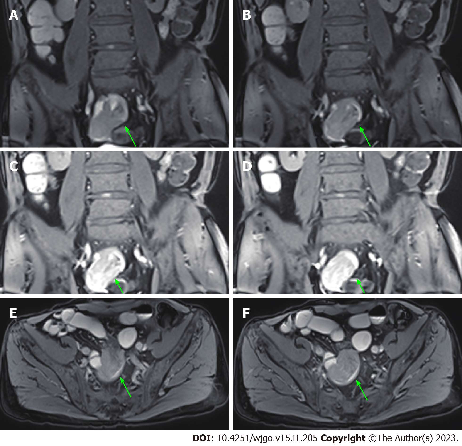
Figure 6 Magnetic resonance enterography after the third admission.
A soft tissue mass (24 mm × 21 mm) (green arrows) was detected in the small intestine near the right pelvis, indicating small intestinal tumor with intussusception. A-D: Representative coronal views; E and F: Representative axial views.
- Citation: Fan WJ, Cheng HH, Wei W. Surgical treatments of recurrent small intestine metastatic melanoma manifesting with gastrointestinal hemorrhage and intussusception: A case report. World J Gastrointest Oncol 2023; 15(1): 205-214
- URL: https://www.wjgnet.com/1948-5204/full/v15/i1/205.htm
- DOI: https://dx.doi.org/10.4251/wjgo.v15.i1.205


