Copyright
©The Author(s) 2023.
World J Gastrointest Oncol. Jan 15, 2023; 15(1): 195-204
Published online Jan 15, 2023. doi: 10.4251/wjgo.v15.i1.195
Published online Jan 15, 2023. doi: 10.4251/wjgo.v15.i1.195
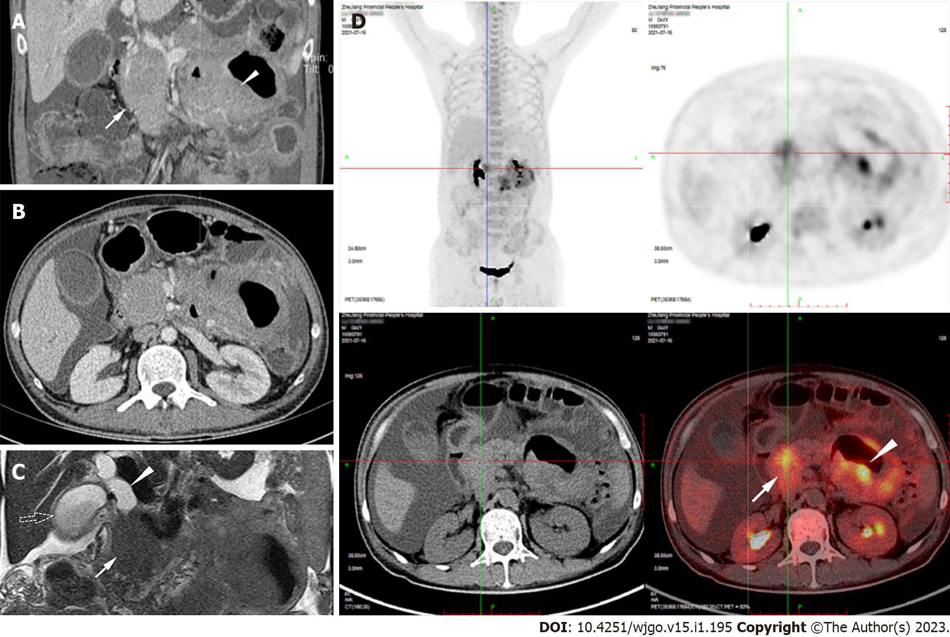
Figure 1 Radiology findings of the lesions.
A: There was circular solid occupying lesion in pancreatic head with 88 mm × 47 mm × 52 mm in size (white arrow). Jejunal wall thickened to 34 mm (white arrow head), without intestinal stenosis and obstructive signs; B: These two lesions were both in uniform computed tomography (CT) density and moderate enhancement; C: Magnetic resonance cholangiopancreatography revealed the head pancreatic mass (white arrow) with dilated biliary duct (white arrow head) and enlarged gallbladder (dotted arrow); D: 18F-Fluorodeoxyglucose(18-F FDG) positron emission tomography/CT detected intensive FDG uptake within the pancreatic head lesion (white arrow) and the jejunal wall thickening (white arrow head).
- Citation: Wang YN, Zhu YM, Lei XJ, Chen Y, Ni WM, Fu ZW, Pan WS. Intestinal natural killer/T-cell lymphoma presenting as a pancreatic head space-occupying lesion: A case report. World J Gastrointest Oncol 2023; 15(1): 195-204
- URL: https://www.wjgnet.com/1948-5204/full/v15/i1/195.htm
- DOI: https://dx.doi.org/10.4251/wjgo.v15.i1.195


