Copyright
©The Author(s) 2023.
World J Gastrointest Oncol. Jan 15, 2023; 15(1): 186-194
Published online Jan 15, 2023. doi: 10.4251/wjgo.v15.i1.186
Published online Jan 15, 2023. doi: 10.4251/wjgo.v15.i1.186
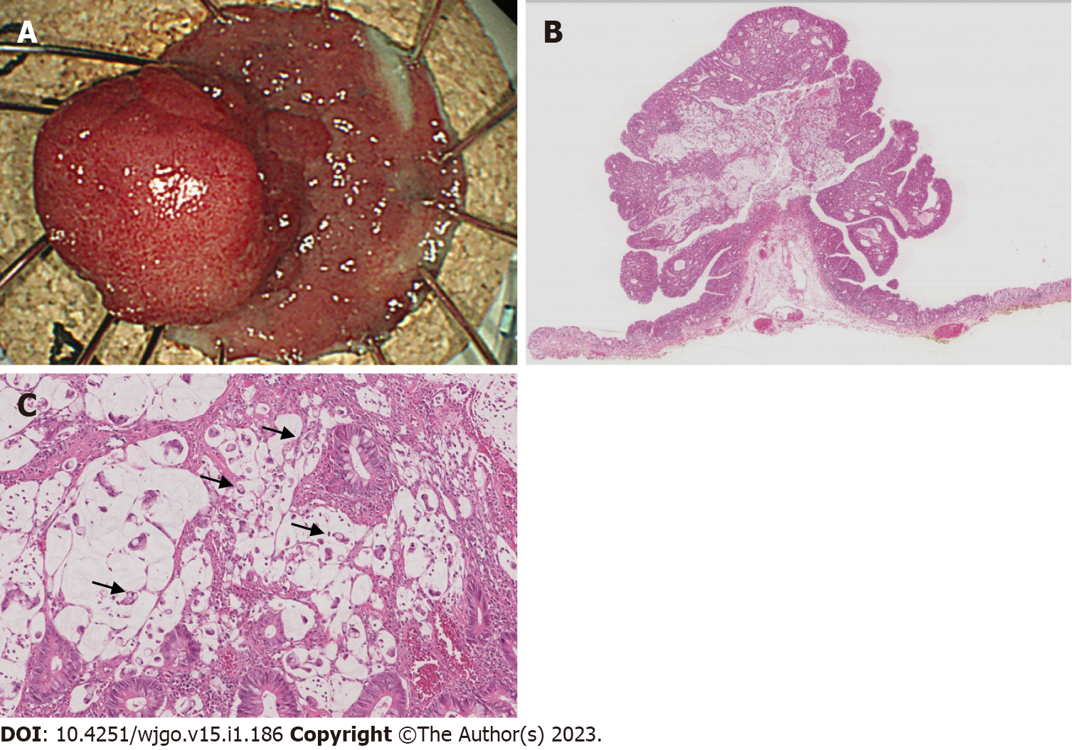
Figure 3 A histopathological examination of the endoscopically resected specimen.
A: A protruded polyp and flat elevation are removed by endoscopic submucosal dissection. The tumor margin is surrounded by normal epithelia, indicating R0 resection; B: Adenocarcinoma composed of mucinous and tubular carcinoma with an adenoma component; C: Signet ring cell carcinoma is observed in mucinous lake (arrows).
- Citation: Murakami Y, Tanabe H, Ono Y, Sugiyama Y, Kobayashi Y, Kunogi T, Sasaki T, Takahashi K, Ando K, Ueno N, Kashima S, Yuzawa S, Moriichi K, Mizukami Y, Fujiya M, Okumura T. Local recurrence after successful endoscopic submucosal dissection for rectal mucinous mucosal adenocarcinoma: A case report. World J Gastrointest Oncol 2023; 15(1): 186-194
- URL: https://www.wjgnet.com/1948-5204/full/v15/i1/186.htm
- DOI: https://dx.doi.org/10.4251/wjgo.v15.i1.186


