Copyright
©The Author(s) 2023.
World J Gastrointest Oncol. Jan 15, 2023; 15(1): 186-194
Published online Jan 15, 2023. doi: 10.4251/wjgo.v15.i1.186
Published online Jan 15, 2023. doi: 10.4251/wjgo.v15.i1.186
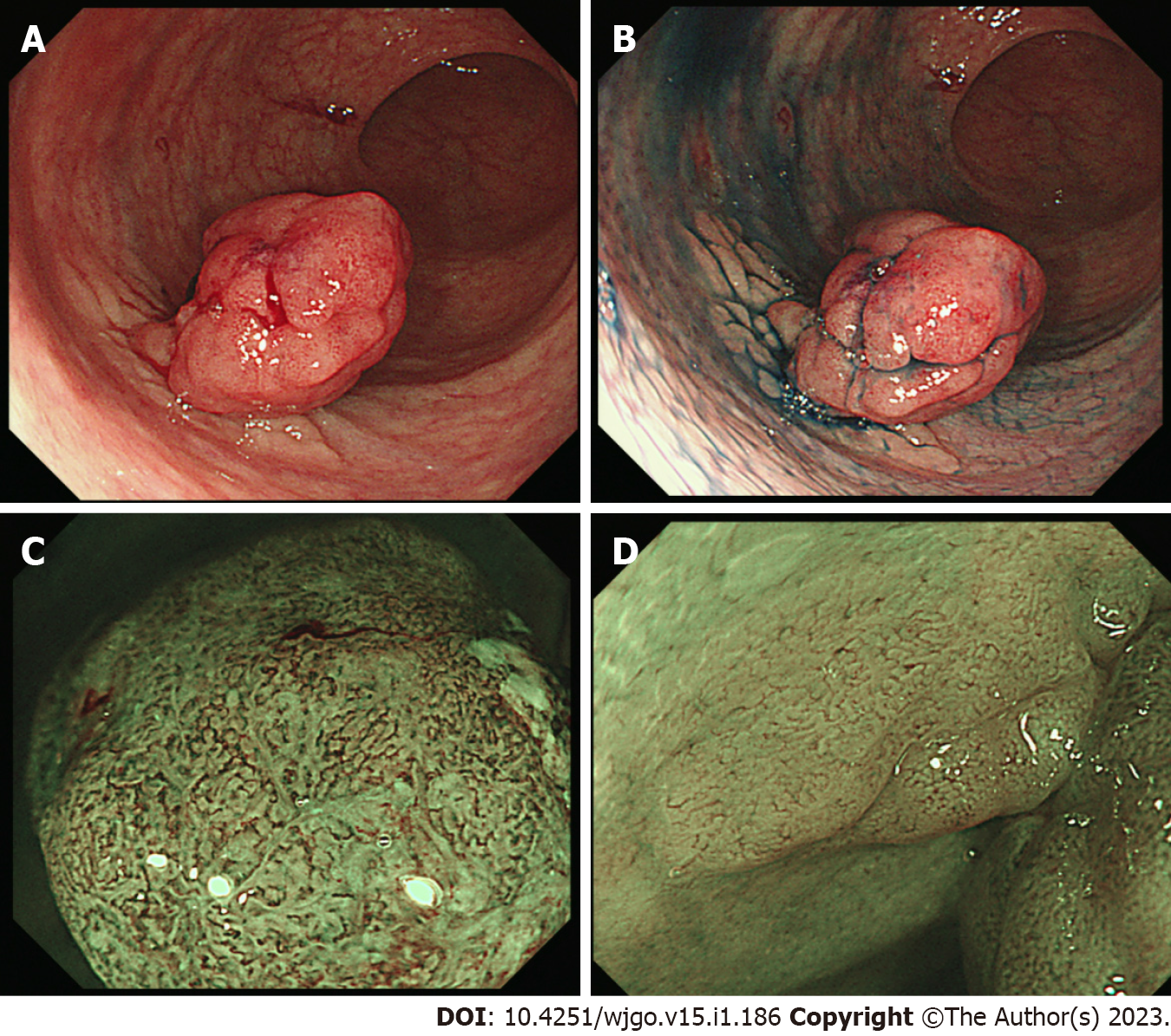
Figure 1 Endoscopic findings of the rectal tumor.
A: A remarkable protrusion (Is) with slight bleeding is observed in the rectum; B: Chromoendoscopy enhances a flat elevated lesion (IIa) which is located at the base of the protrusion lesion; C: Magnified endoscopy with narrow-band imaging reveals an intense irregular micro-vascular pattern indicating the existence of carcinoma in the Is lesion; D: Magnified endoscopy shows faint vascular pattern on the IIa lesion.
- Citation: Murakami Y, Tanabe H, Ono Y, Sugiyama Y, Kobayashi Y, Kunogi T, Sasaki T, Takahashi K, Ando K, Ueno N, Kashima S, Yuzawa S, Moriichi K, Mizukami Y, Fujiya M, Okumura T. Local recurrence after successful endoscopic submucosal dissection for rectal mucinous mucosal adenocarcinoma: A case report. World J Gastrointest Oncol 2023; 15(1): 186-194
- URL: https://www.wjgnet.com/1948-5204/full/v15/i1/186.htm
- DOI: https://dx.doi.org/10.4251/wjgo.v15.i1.186


