Copyright
©The Author(s) 2022.
World J Gastrointest Oncol. Sep 15, 2022; 14(9): 1808-1822
Published online Sep 15, 2022. doi: 10.4251/wjgo.v14.i9.1808
Published online Sep 15, 2022. doi: 10.4251/wjgo.v14.i9.1808
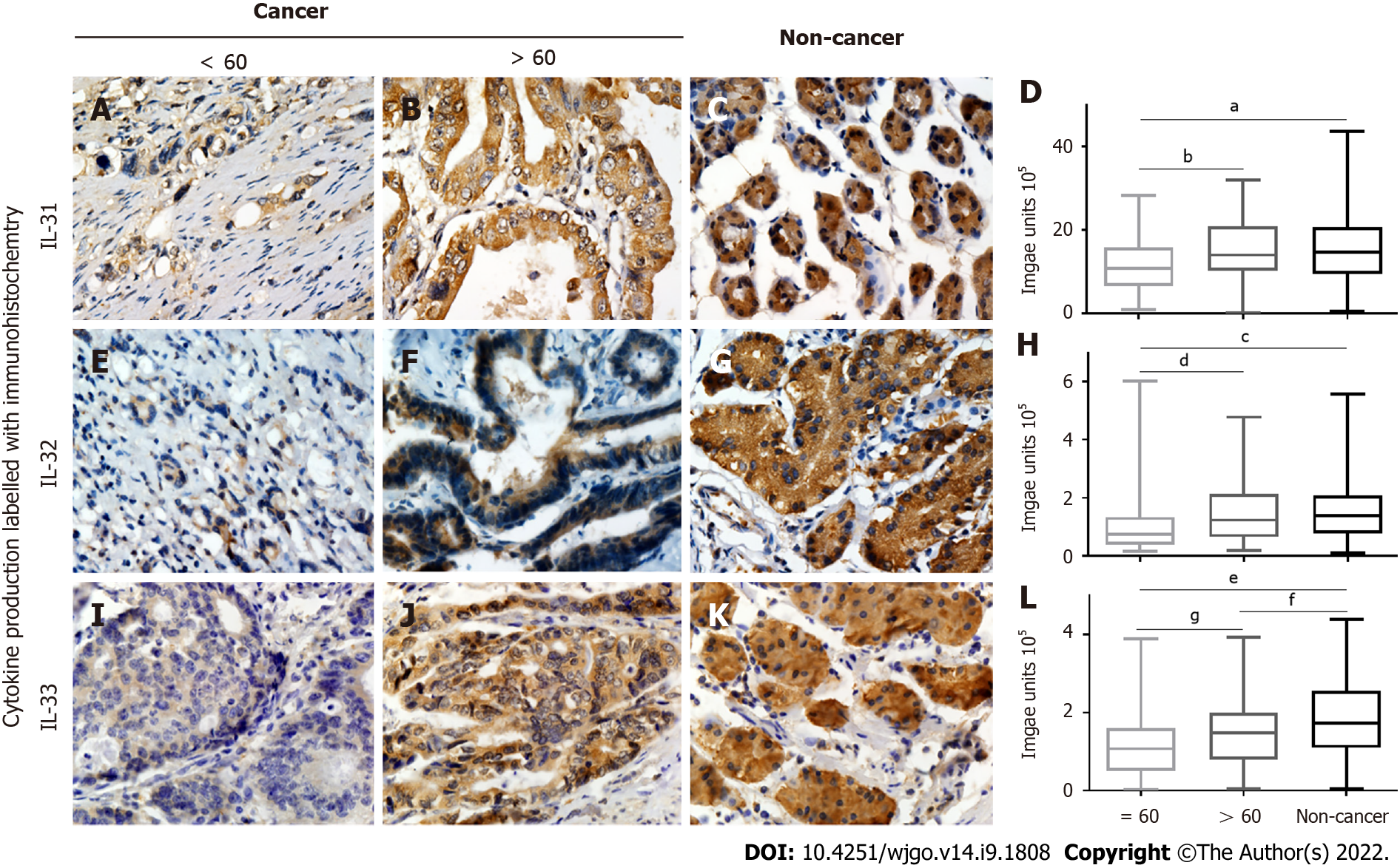
Figure 3 Correlation of interleukin-31, interleukin-32, and interleukin-33 expression with age.
A-D: Positive (brown) interleukin (IL)-31 expression in gastric cancer tissues from patients aged < 60 (A) vs > 60 (B) years and noncancerous tissues (C) plus quantified data (D); E-H: Positive (brown) IL-32 expression in gastric cancer tissues from patients aged < 60 (E) vs > 60 (F) years and noncancerous tissues (G), and quantified data (H); I-L: Positive (brown) IL33 expression in gastric cancer tissues from patients aged < 60 (I) vs > 60 (J) years and noncancerous tissues (K) plus quantified data (L). IL-32 and IL-33 were all decreased in the group of gastric cancer patients aged less than or equal to 60 years. aP < 0.01; bP < 0.01; cP < 0.0001; dP < 0.01; eP < 0.0001; fP < 0.05; gP < 0.05. IL: Interleukin.
- Citation: Liu QH, Zhang JW, Xia L, Wise SG, Hambly BD, Tao K, Bao SS. Clinical implications of interleukins-31, 32, and 33 in gastric cancer. World J Gastrointest Oncol 2022; 14(9): 1808-1822
- URL: https://www.wjgnet.com/1948-5204/full/v14/i9/1808.htm
- DOI: https://dx.doi.org/10.4251/wjgo.v14.i9.1808


