Copyright
©The Author(s) 2022.
World J Gastrointest Oncol. Sep 15, 2022; 14(9): 1739-1757
Published online Sep 15, 2022. doi: 10.4251/wjgo.v14.i9.1739
Published online Sep 15, 2022. doi: 10.4251/wjgo.v14.i9.1739
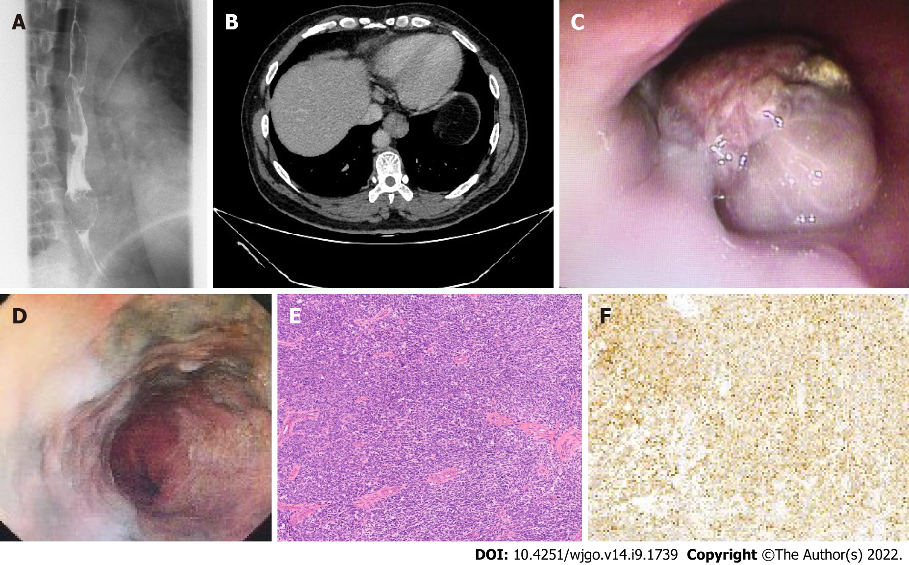
Figure 1 Imaging and microphotograph of primary malignant melanoma of the esophagus.
A: Barium swallow examination showed an irregular filling defect on the lower third of the esophagus, causing mucosa destruction; B: Computed tomography showed an eccentric thickening in the lower third of the esophagus wall, with enhancement; C and D: Esophagoscopy revealed a nonpigmented polypoid tumor with hyperemia and erosion in the lower esophagus, and black lesion scattered on the wall of esophagus; E: Hematoxylin-eosin staining identified malignant melanoma cells in the lamina propria of the esophagus (× 100); F: Immunohistochemical staining with HMB45 (human melanoma black 45) antibody revealed positive tumor cells (× 100).
- Citation: Zhou SL, Zhang LQ, Zhao XK, Wu Y, Liu QY, Li B, Wang JJ, Zhao RJ, Wang XJ, Chen Y, Wang LD, Kong LF. Clinicopathological characterization of ten patients with primary malignant melanoma of the esophagus and literature review. World J Gastrointest Oncol 2022; 14(9): 1739-1757
- URL: https://www.wjgnet.com/1948-5204/full/v14/i9/1739.htm
- DOI: https://dx.doi.org/10.4251/wjgo.v14.i9.1739


