Copyright
©The Author(s) 2022.
World J Gastrointest Oncol. Sep 15, 2022; 14(9): 1622-1636
Published online Sep 15, 2022. doi: 10.4251/wjgo.v14.i9.1622
Published online Sep 15, 2022. doi: 10.4251/wjgo.v14.i9.1622
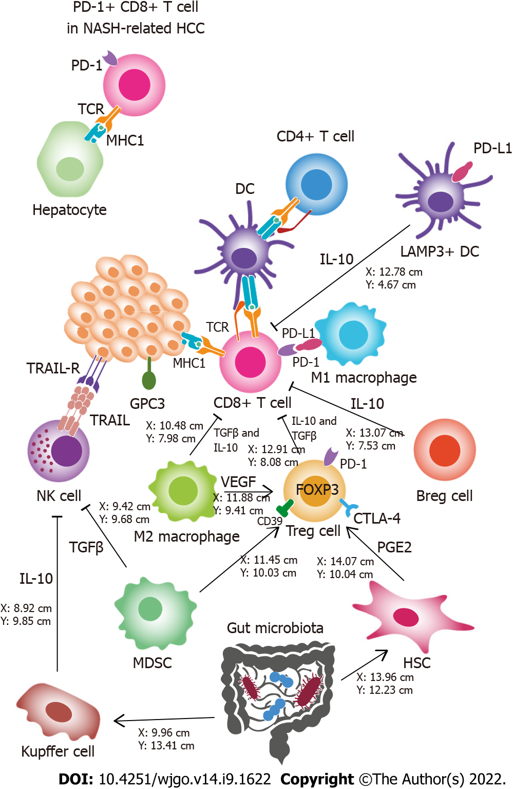
Figure 1 Hepatocellular carcinoma immunological microenvironment.
Different elements that contribute to the antitumor activity or limit antitumor immunity are illustrated schematically. The main effectors against tumor cells are CD8+ T cells and natural killer (NK) cells. Dendritic cells (DCs), CD4+ cells, and M1 macrophages enhance CD8+ T cell cytotoxicity. Regulatory T cells, regulatory B cells, LAMP3+ DCs, and M2 macrophages inhibit CD8+ T cells and induce an immunosuppressive environment. Tumor cells attract M2 macrophages by expressing glypican-3 (GPC3). Myeloid-derived suppressor cells and Kupffer cells produce immunosuppressive cytokines in the tumor microenvironment and inhibit NK cells. The gut microbiota might play an indirect role in immunosuppression through persistent inflammation or other mechanisms leading to immune cell exhaustion. PD-1+ CD8+ T cells in NASH-related hepatocellular carcinoma show cytotoxic activity against hepatocytes, instead of exhibiting antitumor function. Breg: Regulatory B cells; DC: Dendritic cell; HSC: Hepatic stellate cell; MDSC: Myeloid-derived suppressor cell; MHC I: Major histocompatibility complex class I; NASH: Nonalcoholic steatohepatitis; NK: Natural killer; TCR: T cell receptor; TME: Tumor microenvironment; Treg: Regulatory T cells; TRAIL: Tumor necrosis factor-related apoptosis-inducing ligand; TRAIL-R: Tumor necrosis factor-related apoptosis-inducing ligand receptor; VEGF: Vascular endothelial growth factor.
- Citation: Costante F, Airola C, Santopaolo F, Gasbarrini A, Pompili M, Ponziani FR. Immunotherapy for nonalcoholic fatty liver disease-related hepatocellular carcinoma: Lights and shadows. World J Gastrointest Oncol 2022; 14(9): 1622-1636
- URL: https://www.wjgnet.com/1948-5204/full/v14/i9/1622.htm
- DOI: https://dx.doi.org/10.4251/wjgo.v14.i9.1622


