Copyright
©The Author(s) 2022.
World J Gastrointest Oncol. Aug 15, 2022; 14(8): 1585-1593
Published online Aug 15, 2022. doi: 10.4251/wjgo.v14.i8.1585
Published online Aug 15, 2022. doi: 10.4251/wjgo.v14.i8.1585
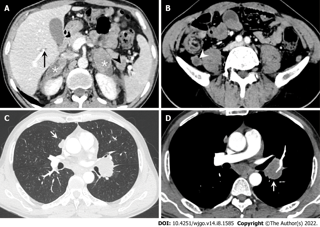
Figure 2 Abdominal and chest computed tomography.
A: Multiple metastatic lesions are observed on the bilateral adrenal gland (*), liver (black arrow) and pancreas (black arrowhead); B: Several enlarged lymph nodes (white arrowhead) are seen on the retroperitoneal area; C: A pulmonary metastatic nodule (short white arrow) is seen in lung windows; D: Several enlarged lymph nodes (short white arrow) are shown on the mediastinum area in contrast-enhanced computed tomography.
- Citation: Guo AW, Liu YS, Li H, Yuan Y, Li SX. Ewing sarcoma of the ileum with wide multiorgan metastases: A case report and review of literature. World J Gastrointest Oncol 2022; 14(8): 1585-1593
- URL: https://www.wjgnet.com/1948-5204/full/v14/i8/1585.htm
- DOI: https://dx.doi.org/10.4251/wjgo.v14.i8.1585


