Copyright
©The Author(s) 2022.
World J Gastrointest Oncol. Aug 15, 2022; 14(8): 1585-1593
Published online Aug 15, 2022. doi: 10.4251/wjgo.v14.i8.1585
Published online Aug 15, 2022. doi: 10.4251/wjgo.v14.i8.1585
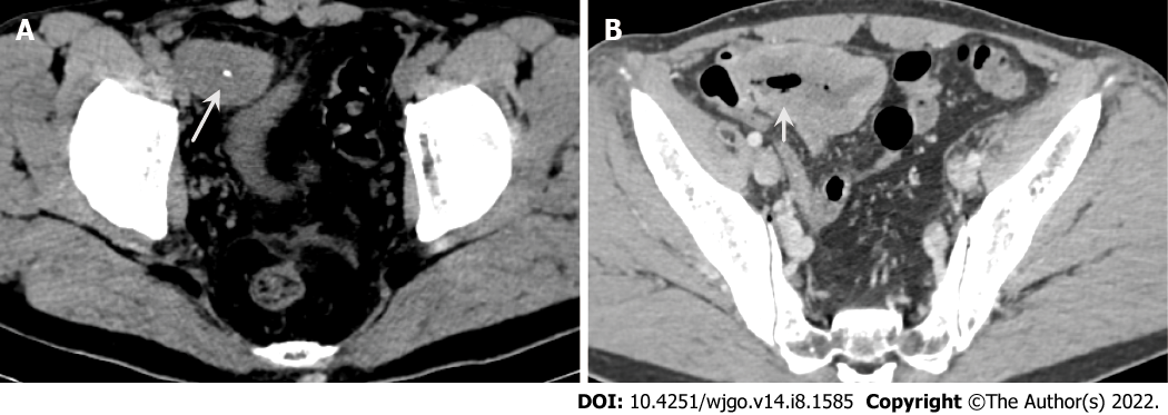
Figure 1 Abdominal computed tomography.
A: Axial computed tomography (CT) image shows a heterogenetic mass with calcification (white arrows); B: Contrast-enhanced CT shows mild heterogenetic enhancement and communication with the small intestinal lumen (short white arrows).
- Citation: Guo AW, Liu YS, Li H, Yuan Y, Li SX. Ewing sarcoma of the ileum with wide multiorgan metastases: A case report and review of literature. World J Gastrointest Oncol 2022; 14(8): 1585-1593
- URL: https://www.wjgnet.com/1948-5204/full/v14/i8/1585.htm
- DOI: https://dx.doi.org/10.4251/wjgo.v14.i8.1585


