Copyright
©The Author(s) 2022.
World J Gastrointest Oncol. Jul 15, 2022; 14(7): 1363-1371
Published online Jul 15, 2022. doi: 10.4251/wjgo.v14.i7.1363
Published online Jul 15, 2022. doi: 10.4251/wjgo.v14.i7.1363
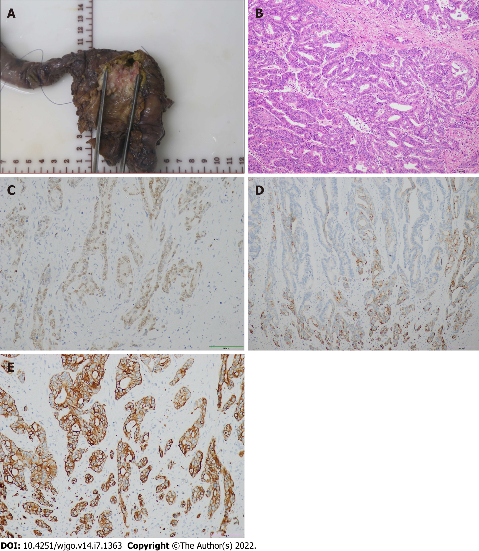
Figure 2 Postoperative pathological picture.
A: The mass in the distal common bile duct infiltrated the duodenal wall; B: Well-differentiated adenocarcinoma; C: Immunohistochemical CDX-2 positive; D: Immunohistochemical cytokeratin (CK)7 positive; E: Immunohistochemical CK19 positive.
- Citation: Li BB, Lu SL, He X, Lei B, Yao JN, Feng SC, Yu SP. Da Vinci robot-assisted pancreato-duodenectomy in a patient with situs inversus totalis: A case report and review of literature. World J Gastrointest Oncol 2022; 14(7): 1363-1371
- URL: https://www.wjgnet.com/1948-5204/full/v14/i7/1363.htm
- DOI: https://dx.doi.org/10.4251/wjgo.v14.i7.1363


