Copyright
©The Author(s) 2022.
World J Gastrointest Oncol. May 15, 2022; 14(5): 1057-1064
Published online May 15, 2022. doi: 10.4251/wjgo.v14.i5.1057
Published online May 15, 2022. doi: 10.4251/wjgo.v14.i5.1057
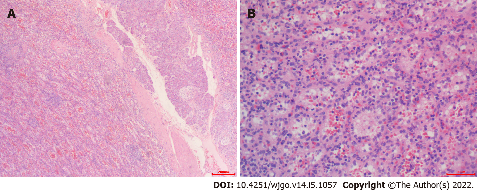
Figure 4 Microscopic examination.
A: The mass grew from the pancreas [hematoxylin and eosin (HE), × 40]; B: The mass contained unorganized small slit-like vascular channels enclosing red blood cells and lined with plump endothelial cells. No area of cytologic atypia was identified. Focal lymphoid aggregates were found in the intravascular areas (HE, × 200). White pulp or fibrosis was not observed.
- Citation: Xu SY, Zhou B, Wei SM, Zhao YN, Yan S. Successful treatment of pancreatic accessory splenic hamartoma by laparoscopic spleen-preserving distal pancreatectomy: A case report. World J Gastrointest Oncol 2022; 14(5): 1057-1064
- URL: https://www.wjgnet.com/1948-5204/full/v14/i5/1057.htm
- DOI: https://dx.doi.org/10.4251/wjgo.v14.i5.1057


