Copyright
©The Author(s) 2022.
World J Gastrointest Oncol. May 15, 2022; 14(5): 1037-1049
Published online May 15, 2022. doi: 10.4251/wjgo.v14.i5.1037
Published online May 15, 2022. doi: 10.4251/wjgo.v14.i5.1037
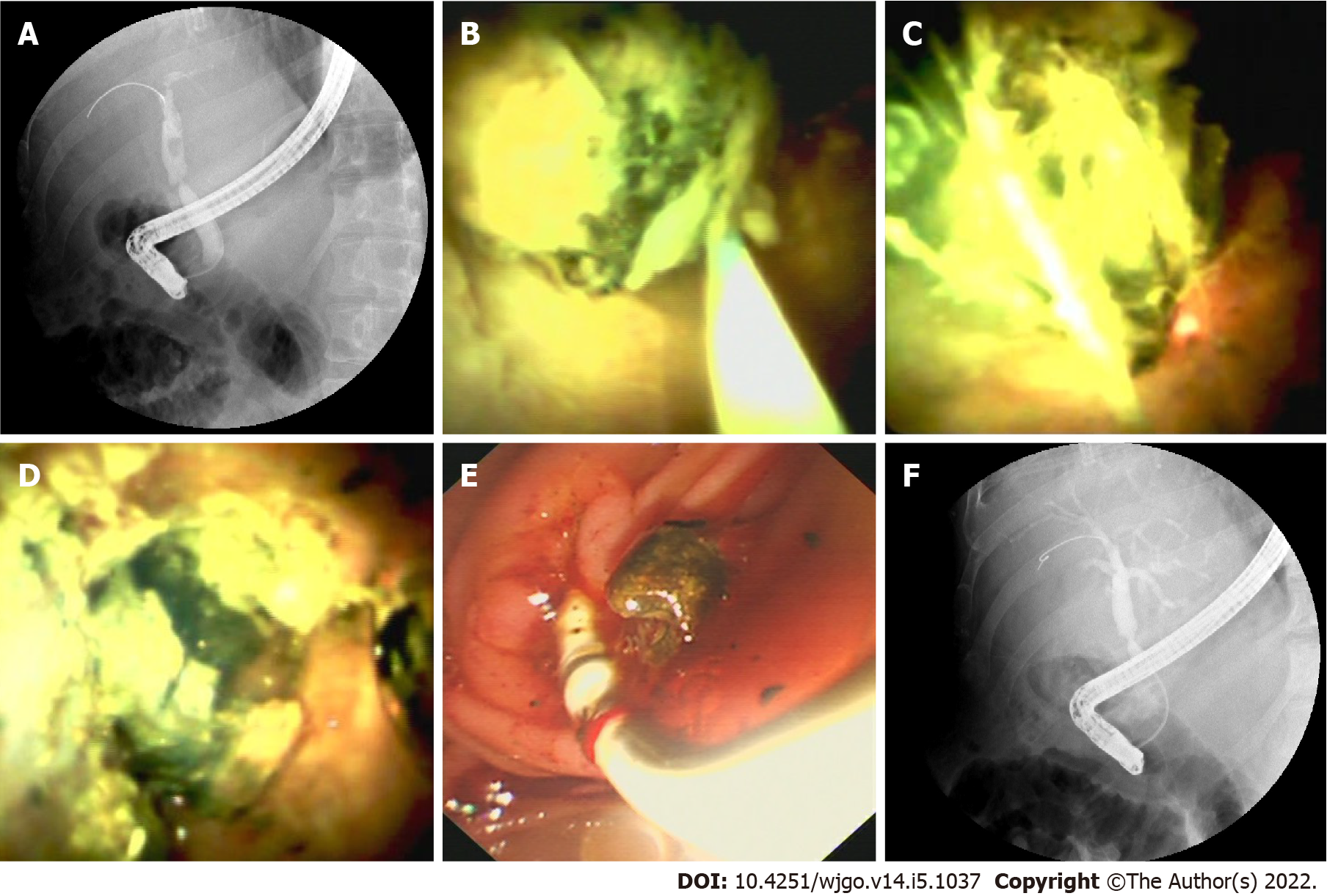
Figure 5 Laser lithotripsy under direct vision.
A: Endoscopic retrograde cholangiopancreatography (ERCP) image shows a stone in the bile duct above an anastomotic stricture; B: Cholangioscopic image shows a green stone in the donor bile duct; C: Optical fiber inserted through the cholangioscopy channel touching the stone; D: Cholangioscopic image shows the stone was shattered by laser; E: Duodenoscopic image shows crushed stone taken out by balloon; F: ERCP image shows the stone was completely extracted.
- Citation: Yu JF, Zhang DL, Wang YB, Hao JY. Digital single-operator cholangioscopy for biliary stricture after cadaveric liver transplantation. World J Gastrointest Oncol 2022; 14(5): 1037-1049
- URL: https://www.wjgnet.com/1948-5204/full/v14/i5/1037.htm
- DOI: https://dx.doi.org/10.4251/wjgo.v14.i5.1037


