Copyright
©The Author(s) 2022.
World J Gastrointest Oncol. May 15, 2022; 14(5): 1037-1049
Published online May 15, 2022. doi: 10.4251/wjgo.v14.i5.1037
Published online May 15, 2022. doi: 10.4251/wjgo.v14.i5.1037
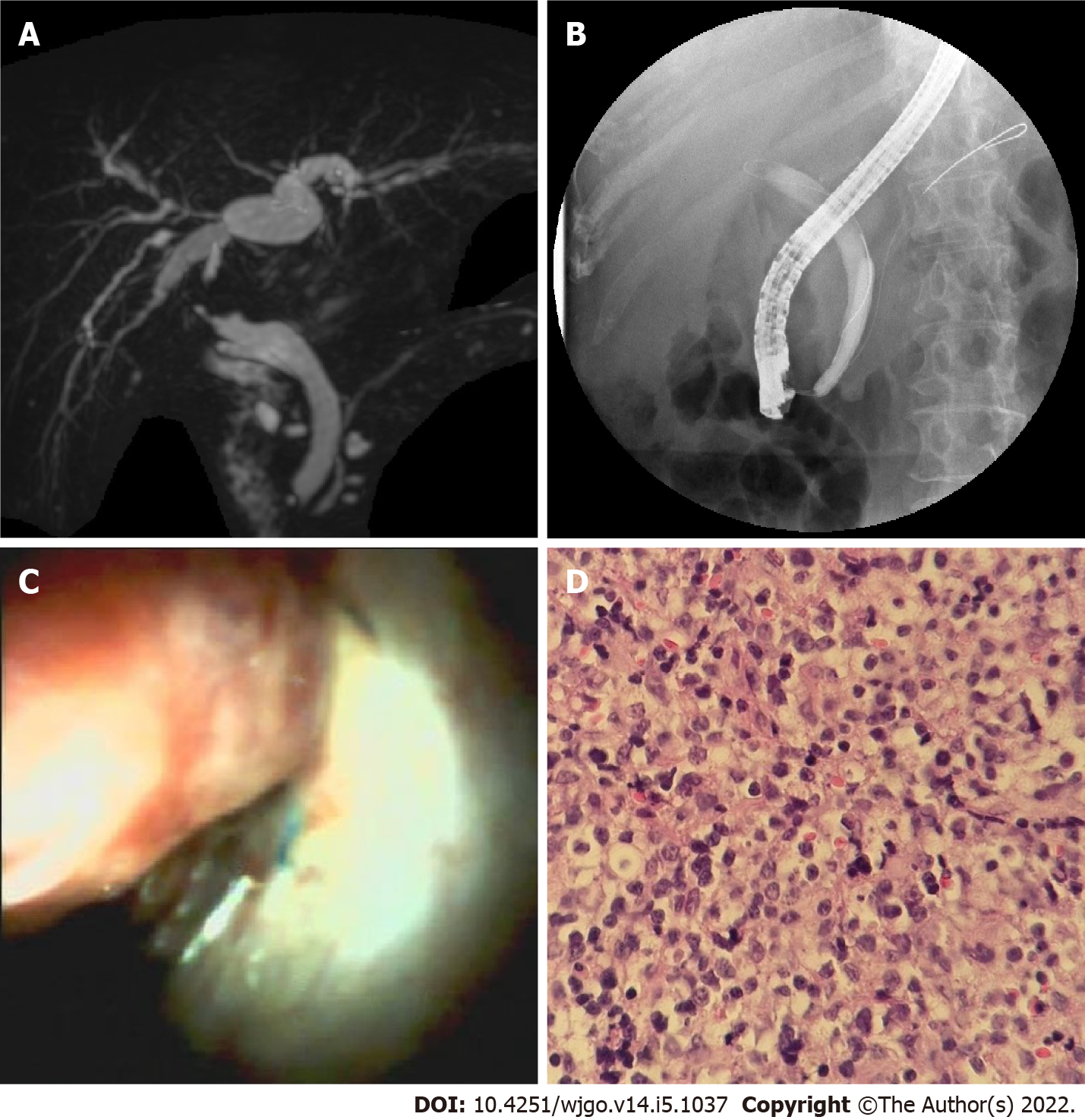
Figure 4 A neoplasm in donor bile duct.
A: Magnetic resonance cholangiopancreatography image shows stricture between anastomosis and hilus region; B: Endoscopic retrograde cholangiopancreatography image shows guidewire failed to pass through the stricture; C: Cholangioscopic image shows the neoplasm with a red surface in the donor bile tract; D: Pathological sections of the neoplasm show many identical lymphocytes with distinct atypia.
- Citation: Yu JF, Zhang DL, Wang YB, Hao JY. Digital single-operator cholangioscopy for biliary stricture after cadaveric liver transplantation. World J Gastrointest Oncol 2022; 14(5): 1037-1049
- URL: https://www.wjgnet.com/1948-5204/full/v14/i5/1037.htm
- DOI: https://dx.doi.org/10.4251/wjgo.v14.i5.1037


