Copyright
©The Author(s) 2022.
World J Gastrointest Oncol. Apr 15, 2022; 14(4): 858-871
Published online Apr 15, 2022. doi: 10.4251/wjgo.v14.i4.858
Published online Apr 15, 2022. doi: 10.4251/wjgo.v14.i4.858
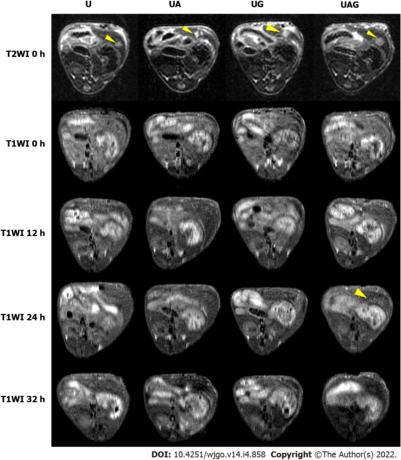
Figure 3 T1-weighted images of hepatocellular carcinoma-model mice administered with ultra-small superparamagnetic iron oxide probes at various time points.
Columns represent livers treated with different probes, and the first row shows pre-injection (0 h) T2-weighted images as references for the tumor location indicated by yellow arrowhead. UA: Anti-AFP-USPIO; UG: Anti-GPC3-USPIO; UAG: Anti-AFP-USPIO-anti-GPC3.
- Citation: Ma XH, Chen K, Wang S, Liu SY, Li DF, Mi YT, Wu ZY, Qu CF, Zhao XM. Bi-specific T1 positive-contrast-enhanced magnetic resonance imaging molecular probe for hepatocellular carcinoma in an orthotopic mouse model. World J Gastrointest Oncol 2022; 14(4): 858-871
- URL: https://www.wjgnet.com/1948-5204/full/v14/i4/858.htm
- DOI: https://dx.doi.org/10.4251/wjgo.v14.i4.858


