Copyright
©The Author(s) 2022.
World J Gastrointest Oncol. Apr 15, 2022; 14(4): 858-871
Published online Apr 15, 2022. doi: 10.4251/wjgo.v14.i4.858
Published online Apr 15, 2022. doi: 10.4251/wjgo.v14.i4.858
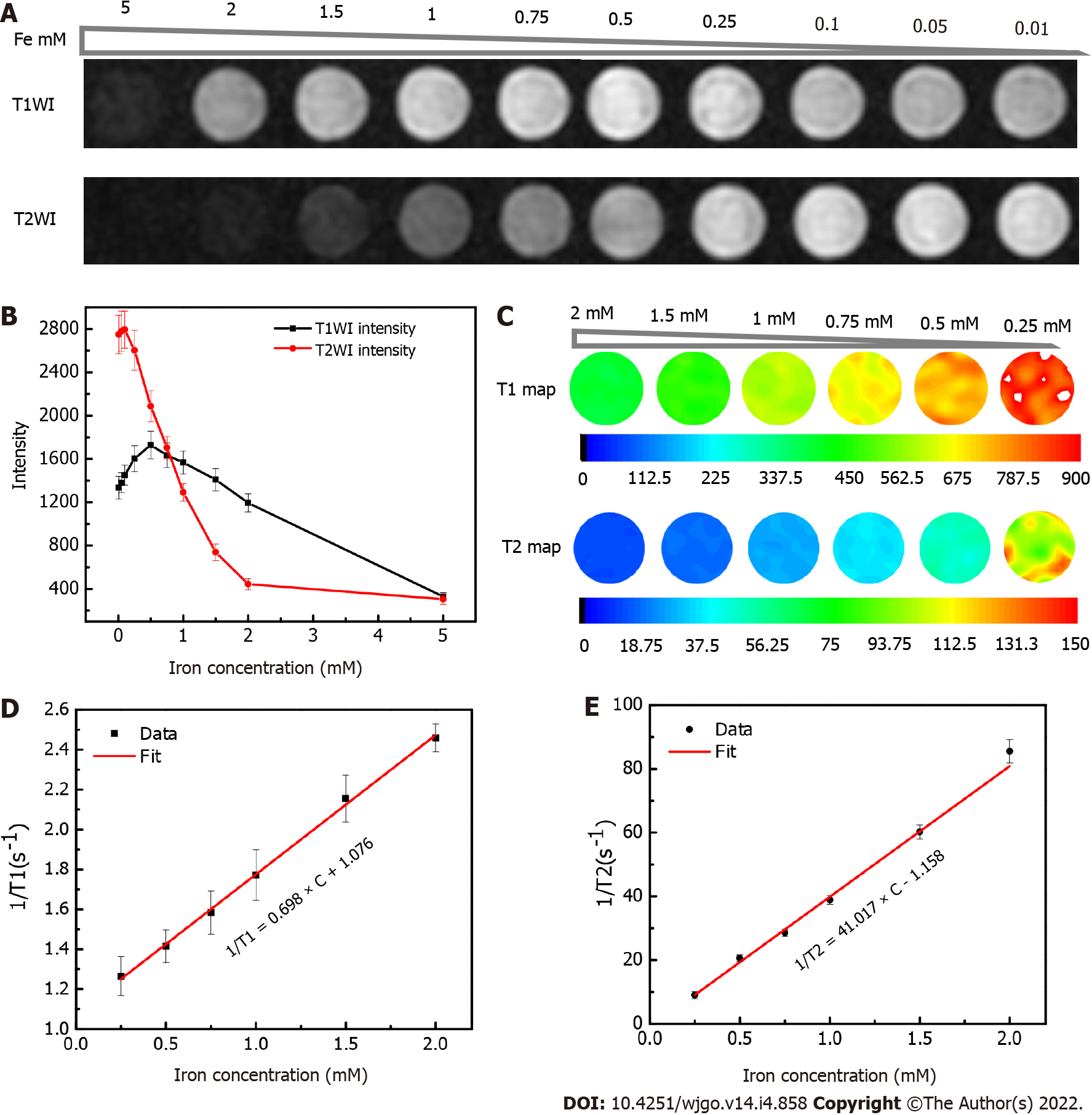
Figure 2 Magnetic resonance imaging properties of ultra-small superparamagnetic iron oxide phantoms.
A: T1- and T2-weighted images of a series of 0.9% saline water solutions containing different concentrations of the ultra-small superparamagnetic iron oxide probes as indicated by iron concentration; B: Changes in the T1- and T2-weighted signal intensities according to iron concentration, with standard deviation also illustrated; C: T1 and T2 map illustrated in pseudo color under different iron concentration; D: Linear regression fitting of the longitudinal relaxation-rate (1/T1); E: transversal relaxation-rate (1/T2) data vs different iron concentrations (with standard deviation also illustrated) for extracting the longitudinal relaxivity (r1) and transverse relaxivity (r2), respectively.
- Citation: Ma XH, Chen K, Wang S, Liu SY, Li DF, Mi YT, Wu ZY, Qu CF, Zhao XM. Bi-specific T1 positive-contrast-enhanced magnetic resonance imaging molecular probe for hepatocellular carcinoma in an orthotopic mouse model. World J Gastrointest Oncol 2022; 14(4): 858-871
- URL: https://www.wjgnet.com/1948-5204/full/v14/i4/858.htm
- DOI: https://dx.doi.org/10.4251/wjgo.v14.i4.858


