Copyright
©The Author(s) 2022.
World J Gastrointest Oncol. Mar 15, 2022; 14(3): 646-653
Published online Mar 15, 2022. doi: 10.4251/wjgo.v14.i3.646
Published online Mar 15, 2022. doi: 10.4251/wjgo.v14.i3.646
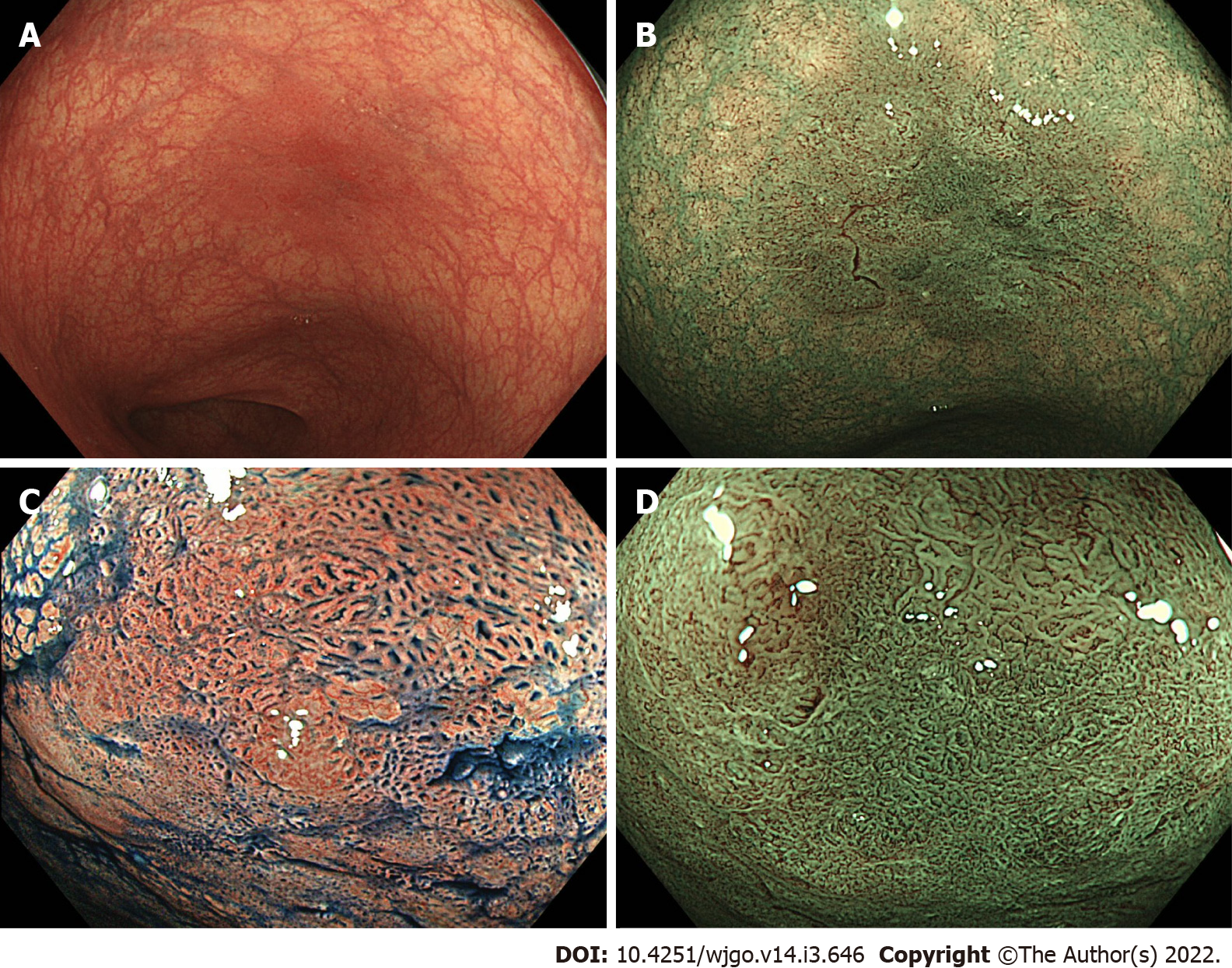
Figure 1 Endoscopic images of a dysplastic lesion in a patient with ulcerative colitis.
A: High-definition colonoscopy with white light shows a tumor recognized by a demarcated, red colored area (Paris classification Type 0-IIa, size 10 mm); B: High-definition colonoscopy with narrow band imaging (NBI); C: Magnifying chromoendoscopy with indigo carmine shows Kudo’s pit pattern types IIIL and VI low-irregularity; D: Magnifying colonoscopy with NBI shows an irregular surface pattern with increased irregular vessels (Type 2B of Japan NBI expert team classification).
- Citation: Akiyama S, Sakamoto T, Steinberg JM, Saito Y, Tsuchiya K. Evolving roles of magnifying endoscopy and endoscopic resection for neoplasia in inflammatory bowel diseases. World J Gastrointest Oncol 2022; 14(3): 646-653
- URL: https://www.wjgnet.com/1948-5204/full/v14/i3/646.htm
- DOI: https://dx.doi.org/10.4251/wjgo.v14.i3.646


