Copyright
©The Author(s) 2022.
World J Gastrointest Oncol. Dec 15, 2022; 14(12): 2415-2421
Published online Dec 15, 2022. doi: 10.4251/wjgo.v14.i12.2415
Published online Dec 15, 2022. doi: 10.4251/wjgo.v14.i12.2415
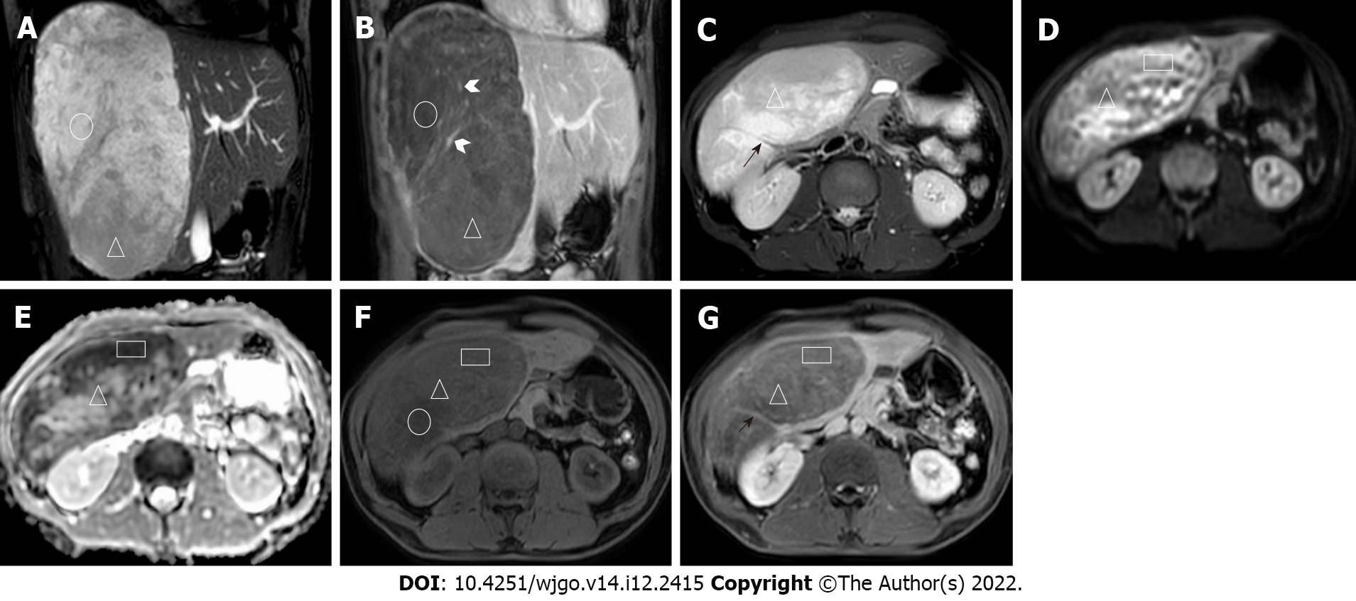
Figure 2 Magnetic resonance imaging images.
A: Coronal balanced fast field echo (B-FFE) image; B: Coronal T1-weighted image (T1WI) after 150-s gadolinium enhancement; C: Axial image, including T2-weighted imaging (T2WI); D: Diffusion-weighted imaging (DWI); E: Apparent diffusion coefficient (ADC); F: T1WI; G: After 7-min gadolinium enhancement, the lesions showed a high signal on B-FFE, a low signal on T1WI, and no definitive enhancement (circled areas). In the lesions, a strip-shaped flow void vascular signal (black short arrow in Figure 2C) can be seen on T2WI. Multiple irregular strip-like obvious enhancement foci with irregular shapes are shown (white arrowhead in Figure 2B). Some areas of high signal on DWI and low signal on ADC and B-FFE (triangular areas) showing progressive and heterogeneous enhancement with gadolinium contrast enhancement. The mass resembled different meteorites in the various sequences.
- Citation: Li DF, Guo XJ, Song SP, Li HB. Rare massive hepatic hemangioblastoma: A case report. World J Gastrointest Oncol 2022; 14(12): 2415-2421
- URL: https://www.wjgnet.com/1948-5204/full/v14/i12/2415.htm
- DOI: https://dx.doi.org/10.4251/wjgo.v14.i12.2415


