Copyright
©The Author(s) 2022.
World J Gastrointest Oncol. Dec 15, 2022; 14(12): 2404-2414
Published online Dec 15, 2022. doi: 10.4251/wjgo.v14.i12.2404
Published online Dec 15, 2022. doi: 10.4251/wjgo.v14.i12.2404
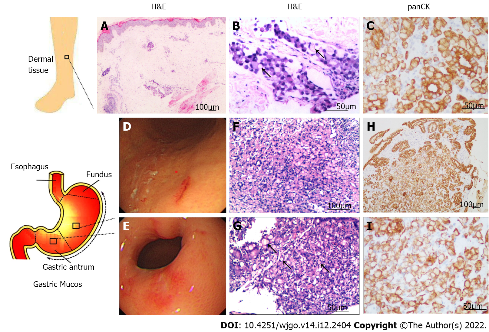
Figure 3 The immunohistochemistry and gastric endoscopy.
A: H&E histological samples of the skin tissue on the right lower limb, 10 ×; B: H&E histological samples of the skin tissue on the right lower limb, 20 ×; C: Histological samples of the skin tissue on the right lower limb stained for panCK, 20 ×; D: Images of gastric endoscopy: Gastric body; E: Images of gastric endoscopy: Gastric antrum; F: H&E histological samples of the mucosa in gastric body, 10 ×; G: Gastric antrum, 20 ×; H: Histological samples of the mucosa in gastric body stained for panCK, 20 ×; I: Gastric antrum, 20 ×. Note the abundant signet-ring cells (black arrows) (B) and (G). H&E: Hematoxylin and eosin; panCK: Pan-cytokeratin.
- Citation: Wang B, Chen J, Wang Y, Dong LL, Shen GF. Edema of limbs as the primary symptom of gastric signet-ring cell carcinoma: A case report and literature review. World J Gastrointest Oncol 2022; 14(12): 2404-2414
- URL: https://www.wjgnet.com/1948-5204/full/v14/i12/2404.htm
- DOI: https://dx.doi.org/10.4251/wjgo.v14.i12.2404


