Copyright
©The Author(s) 2022.
World J Gastrointest Oncol. Nov 15, 2022; 14(11): 2288-2294
Published online Nov 15, 2022. doi: 10.4251/wjgo.v14.i11.2288
Published online Nov 15, 2022. doi: 10.4251/wjgo.v14.i11.2288
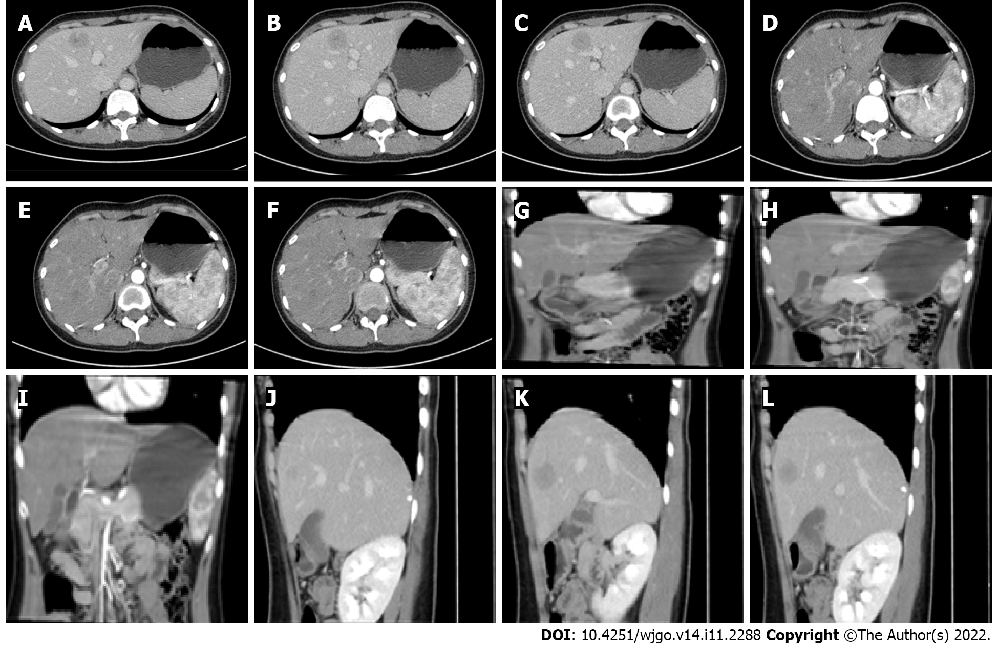
Figure 2 Abdominal contrast-enhanced computed tomography before surgery.
Nodular abnormal enhancement foci (about 27 mm × 26 mm) can be seen in the left inner lobe of the liver, showing uneven and obvious enhancement in the arterial phase and weakening in the venous phase, and a pseudo-capsule can be seen surrounding it. A-F: Axial position; G-I: Coronal position; J-L: Sagittal position.
- Citation: Fu LY, Jiang JL, Liu M, Li JJ, Liu KP, Zhu HT. Surgical treatment of liver inflammatory pseudotumor-like follicular dendritic cell sarcoma: A case report. World J Gastrointest Oncol 2022; 14(11): 2288-2294
- URL: https://www.wjgnet.com/1948-5204/full/v14/i11/2288.htm
- DOI: https://dx.doi.org/10.4251/wjgo.v14.i11.2288


