Copyright
©The Author(s) 2022.
World J Gastrointest Oncol. Nov 15, 2022; 14(11): 2266-2272
Published online Nov 15, 2022. doi: 10.4251/wjgo.v14.i11.2266
Published online Nov 15, 2022. doi: 10.4251/wjgo.v14.i11.2266
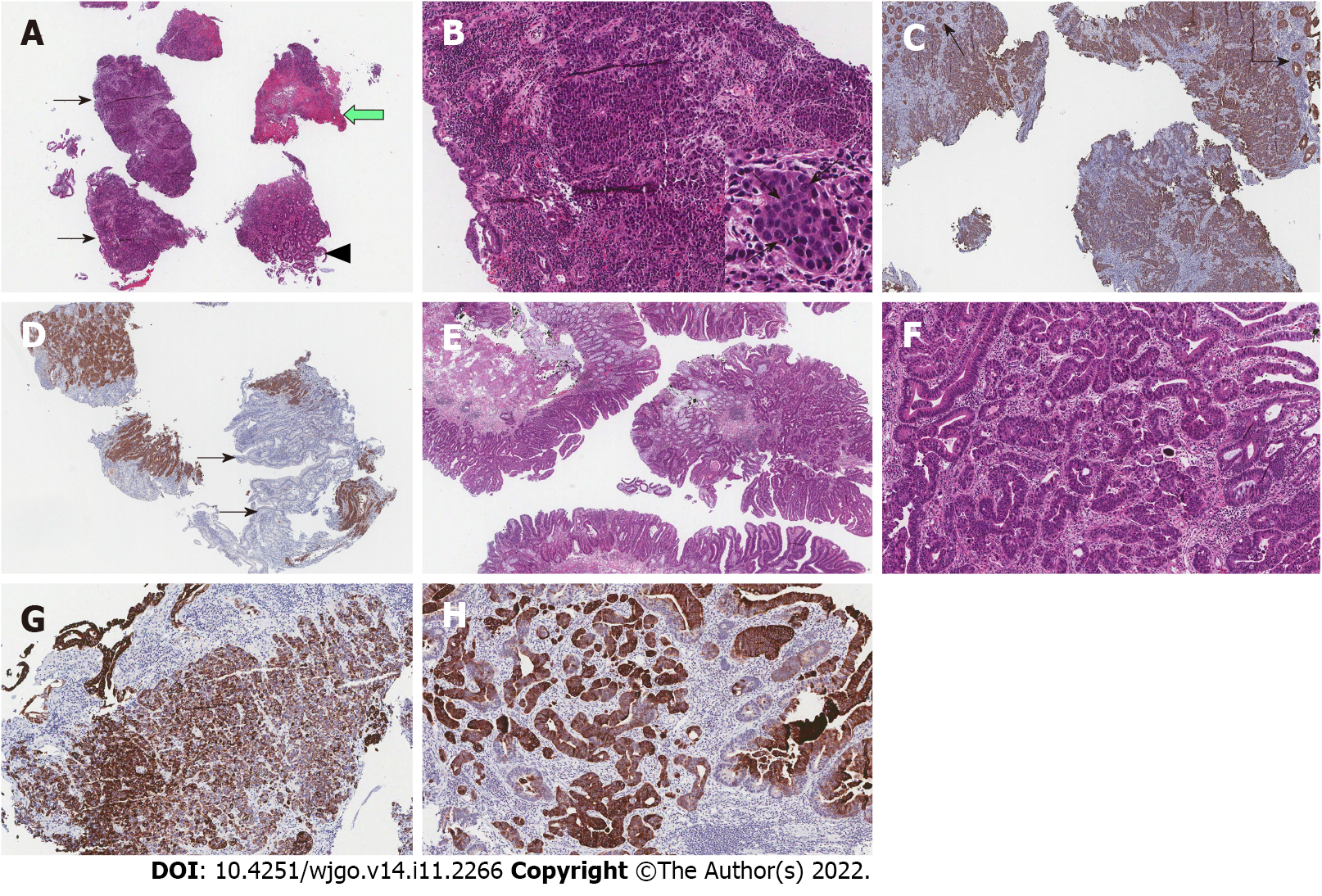
Figure 3 Histologic and immunohistochemical findings of colonic large cell neuroendocrine carcinoma.
Histologic findings of colonic adenocarcinoma. Immunohistochemical stain for CK20 in both colonic large cell neuroendocrine carcinoma and colonic adenocarcinoma. A: Low power view of biopsy specimen reveals several tissue fragments mainly consisted of tumor (black arrows), ulcer debris (white arrow), and non-neoplastic colonic mucosa (arrowhead) (HE, × 20); B: Medium power view displays invasive tumor cells forming organoid structures such as cords or small nests (HE, × 100). Tumor cells display severe atypia and have round nuclei, sometimes with prominent nucleoli (arrows), and moderate amounts of cytoplasm, with high mitotic rate (Inset, HE, × 400); C: Infiltrating tumor cells are identified by immunohistochemical stain for cytokeratin (CK, × 100). Note the non-neoplastic mucosal glands (arrows) that are separated from the tumor; D: Tumor cells show strong immunoreactivity for neuroendocrine marker (synaptophysin, × 100). Note the non-neoplastic mucosal glands (arrows) are negative for neuroendocrine marker; E: Low power view of biopsy specimen reveals colonic epithelial proliferative lesion forming tubular and papillary structures (HE, × 20); F: Medium power view displays invasive growth of tumor cells that form irregular branching and budding of glands (HE, × 100); G: Infiltrating tumor cells of colonic large cell neuroendocrine carcinoma are immunoreactive for CK20 (× 100); H: Invasive tumor glands of colonic adenocarcinoma are also immunoreactive for CK20 (× 100).
- Citation: Baek HS, Kim SW, Lee ST, Park HS, Seo SY. Silent advanced large cell neuroendocrine carcinoma with synchronous adenocarcinoma of the colon: A case report. World J Gastrointest Oncol 2022; 14(11): 2266-2272
- URL: https://www.wjgnet.com/1948-5204/full/v14/i11/2266.htm
- DOI: https://dx.doi.org/10.4251/wjgo.v14.i11.2266


