Copyright
©The Author(s) 2022.
World J Gastrointest Oncol. Nov 15, 2022; 14(11): 2266-2272
Published online Nov 15, 2022. doi: 10.4251/wjgo.v14.i11.2266
Published online Nov 15, 2022. doi: 10.4251/wjgo.v14.i11.2266
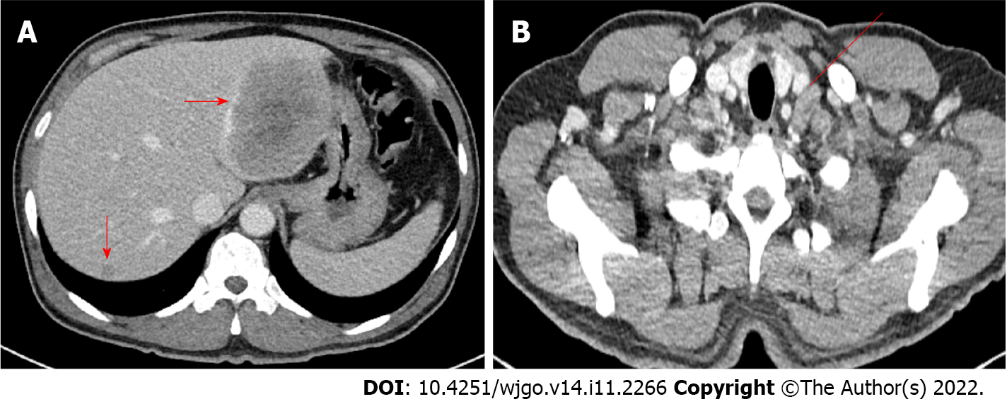
Figure 2 Abdominopelvic computed tomography and chest computed tomography images.
A: Low density lesion of about 8.7 cm in the left lobe of the liver, and several smaller lesions in the right lobe of the liver (arrow); B: Left supraclavicular lymph node enlargement (line).
- Citation: Baek HS, Kim SW, Lee ST, Park HS, Seo SY. Silent advanced large cell neuroendocrine carcinoma with synchronous adenocarcinoma of the colon: A case report. World J Gastrointest Oncol 2022; 14(11): 2266-2272
- URL: https://www.wjgnet.com/1948-5204/full/v14/i11/2266.htm
- DOI: https://dx.doi.org/10.4251/wjgo.v14.i11.2266


