Copyright
©The Author(s) 2022.
World J Gastrointest Oncol. Nov 15, 2022; 14(11): 2253-2265
Published online Nov 15, 2022. doi: 10.4251/wjgo.v14.i11.2253
Published online Nov 15, 2022. doi: 10.4251/wjgo.v14.i11.2253
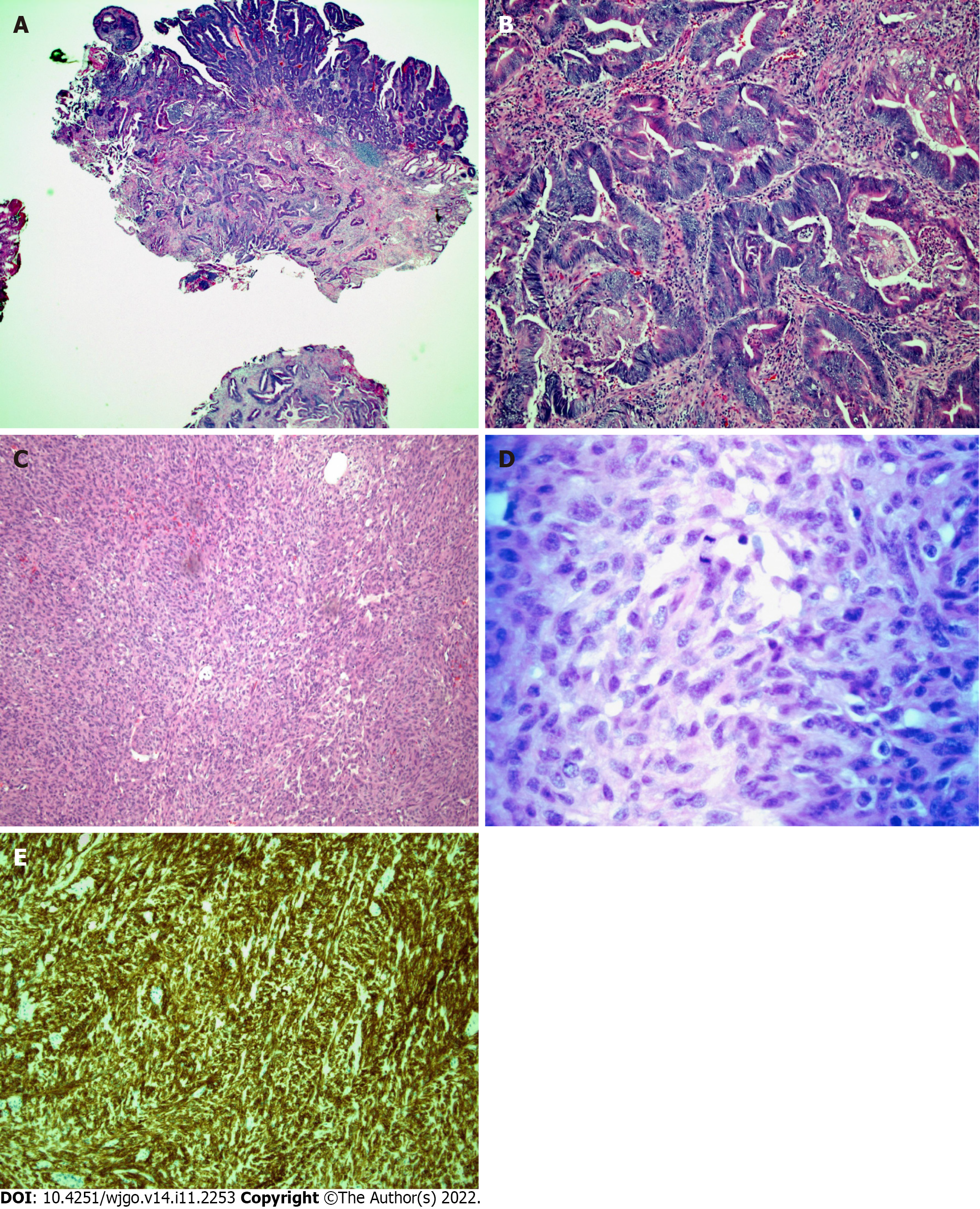
Figure 7 Histopathology of the surgically resected ampullary mass and ileal mass.
A: Low-power micrograph of invasive adenocarcinoma arising from a tubulovillous adenoma with high-grade dysplasia in the ampullary region (hematoxylin and eosin stain, original magnification 20 ×); B: High-power micrograph of ampullary mass showing malignant glands (hematoxylin and eosin stain, original magnification 100 ×); C: Low-power photomicrograph of ileal mass showing the spindle cell morphology of a gastrointestinal stromal tumor (GIST) (hematoxylin and eosin stain, 100 × original magnification); D: A high-power view of cellular mitosis within the ileal GIST (hematoxylin and eosin stain, 600 × original magnification) showed 15 mitoses per 5 square millimeters; E: Immunohistochemical staining for CD117 showing strong and diffuse cytoplasmic immunoreactivity confirming GIST (hematoxylin and eosin stain, 100 × original magnification).
- Citation: Matli VVK, Zibari GB, Wellman G, Ramadas P, Pandit S, Morris J. A rare synchrony of adenocarcinoma of the ampulla with an ileal gastrointestinal stromal tumor: A case report. World J Gastrointest Oncol 2022; 14(11): 2253-2265
- URL: https://www.wjgnet.com/1948-5204/full/v14/i11/2253.htm
- DOI: https://dx.doi.org/10.4251/wjgo.v14.i11.2253


