Copyright
©The Author(s) 2022.
World J Gastrointest Oncol. Nov 15, 2022; 14(11): 2253-2265
Published online Nov 15, 2022. doi: 10.4251/wjgo.v14.i11.2253
Published online Nov 15, 2022. doi: 10.4251/wjgo.v14.i11.2253
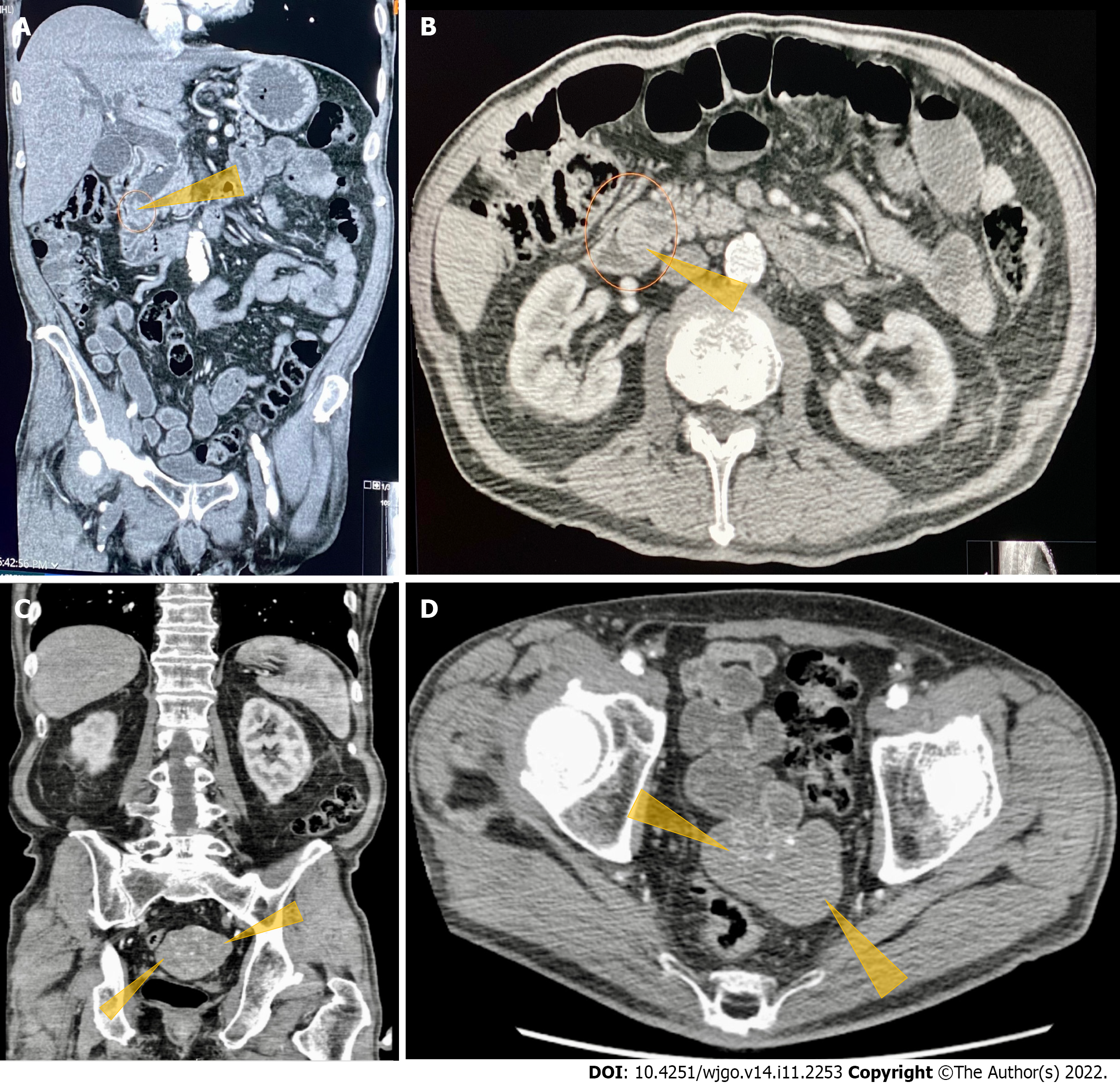
Figure 1 Computerized tomography of the abdomen and pelvis with contrast images.
A: Computerized tomography (CT) abdomen and pelvis with contrast. Coronal section shows obstructed distal common bile duct (CBD) due to a 9 mm polypoid intraluminal (pointed yellow arrows) lesion in the distal CBD; B: CT of the abdomen and pelvis with contrast. Axial section shows polypoid, soft tissue mass at the level of distal common bile duct (pointed yellow arrows); C: CT of the abdomen and pelvis with contrast. Heterogeneously enhanced lobulated mass with punctate calcifications (pointed yellow triangles) in the posterior pelvis originating from the serosal surface of the pelvic small bowel; D: CT of the abdomen and pelvis with contrast. Axial section shows a heterogeneously enhanced lobulated mass with punctate calcifications (pointed yellow triangle) in the posterior pelvis originating from the serosal surface of the pelvic small bowel.
- Citation: Matli VVK, Zibari GB, Wellman G, Ramadas P, Pandit S, Morris J. A rare synchrony of adenocarcinoma of the ampulla with an ileal gastrointestinal stromal tumor: A case report. World J Gastrointest Oncol 2022; 14(11): 2253-2265
- URL: https://www.wjgnet.com/1948-5204/full/v14/i11/2253.htm
- DOI: https://dx.doi.org/10.4251/wjgo.v14.i11.2253


