Copyright
©The Author(s) 2022.
World J Gastrointest Oncol. Oct 15, 2022; 14(10): 2077-2084
Published online Oct 15, 2022. doi: 10.4251/wjgo.v14.i10.2077
Published online Oct 15, 2022. doi: 10.4251/wjgo.v14.i10.2077
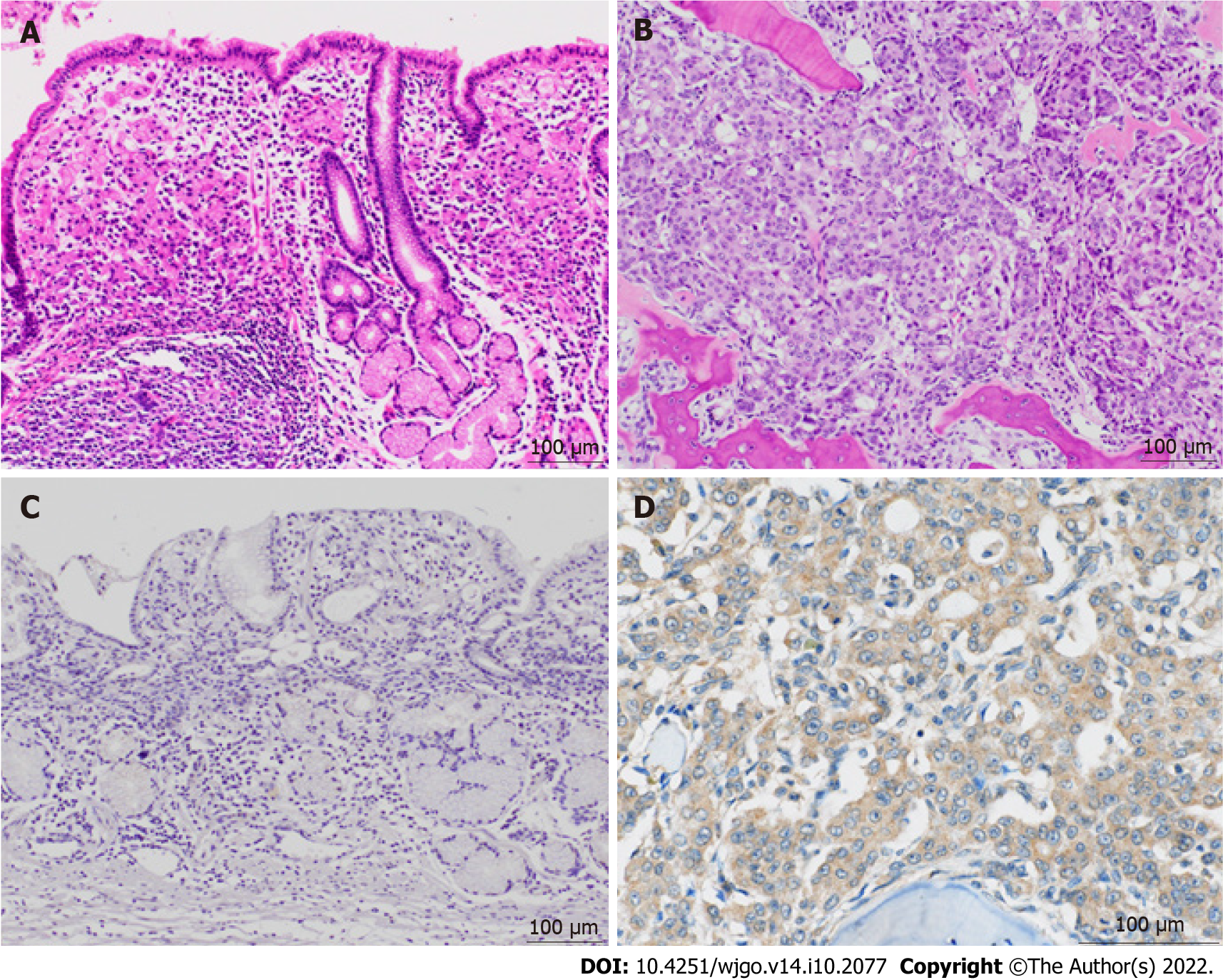
Figure 3 Histologic and immunohistochemical images of the primary and relapsed lesions.
A: Histology of the primary gastric specimen shows moderately to poorly differentiated adenocarcinoma and, partially, signet cell carcinoma (hematoxylin and eosin); B: On autopsy, the metastatic bone marrow lesion shows corresponding adenocarcinoma (hematoxylin and eosin); C: Immunohistochemical staining for granulocyte colony-stimulating factor receptor (G-CSFR) is negative in the primary lesion; D: Immunohistochemical staining for G-CSFR is diffusely positive in the bone marrow metastatic lesion.
- Citation: Fujita K, Okubo A, Nakamura T, Takeuchi N. Disseminated carcinomatosis of the bone marrow caused by granulocyte colony-stimulating factor: A case report and review of literature. World J Gastrointest Oncol 2022; 14(10): 2077-2084
- URL: https://www.wjgnet.com/1948-5204/full/v14/i10/2077.htm
- DOI: https://dx.doi.org/10.4251/wjgo.v14.i10.2077


