Copyright
©The Author(s) 2021.
World J Gastrointest Oncol. Jul 15, 2021; 13(7): 662-672
Published online Jul 15, 2021. doi: 10.4251/wjgo.v13.i7.662
Published online Jul 15, 2021. doi: 10.4251/wjgo.v13.i7.662
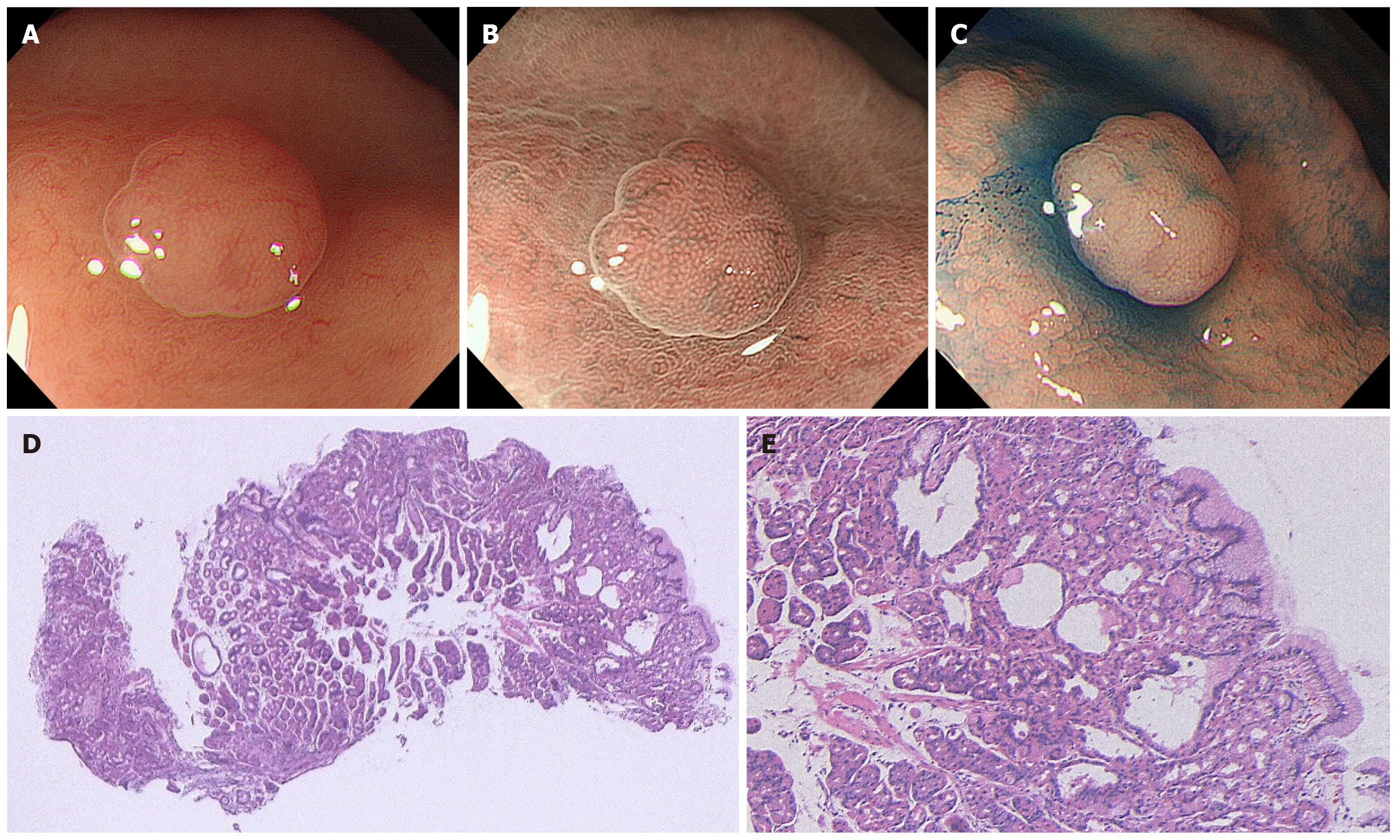
Figure 1 A case of sporadic fundic gland polyp without dysplasia.
A: White light endoscopic view. A 47-year-old man with a complaint of heartburn underwent esophagogastroduodenoscopy. He had no medical history of receiving long-term proton pump inhibitor therapy and no family history of familial adenomatous polyposis (FAP). White light endoscopy identified several fundic gland polyp-like lesions in the body of the stomach without atrophic mucosa, suggesting a Helicobacter pylori-uninfected setting. Among them, a 3 mm isochromatic sessile polyp with a smooth surface structure was detected in the middle body; B: Magnifying narrow-band imaging (NBI) endoscopic view. Magnifying NBI endoscopy revealed regularly arranged, white, dot-shaped crypt openings resembling surrounding mucosa; C: Chromoendoscopic view. Chromoendoscopy using indigo carmine dye revealed less irregularity of the lesion surface. This lesion was endoscopically diagnosed as a fundic gland polyp without dysplasia and was biopsied; D and E: Histopathological view. The histopathological examination revealed a non-dysplastic fundic gland polyp with hyperplastic proliferation of the oxyntic glands and several cystically dilated glands. Colonoscopy did not reveal indications for FAP.
- Citation: Sano W, Inoue F, Hirata D, Iwatate M, Hattori S, Fujita M, Sano Y. Sporadic fundic gland polyps with dysplasia or carcinoma: Clinical and endoscopic characteristics. World J Gastrointest Oncol 2021; 13(7): 662-672
- URL: https://www.wjgnet.com/1948-5204/full/v13/i7/662.htm
- DOI: https://dx.doi.org/10.4251/wjgo.v13.i7.662


