Copyright
©The Author(s) 2021.
World J Gastrointest Oncol. May 15, 2021; 13(5): 409-423
Published online May 15, 2021. doi: 10.4251/wjgo.v13.i5.409
Published online May 15, 2021. doi: 10.4251/wjgo.v13.i5.409
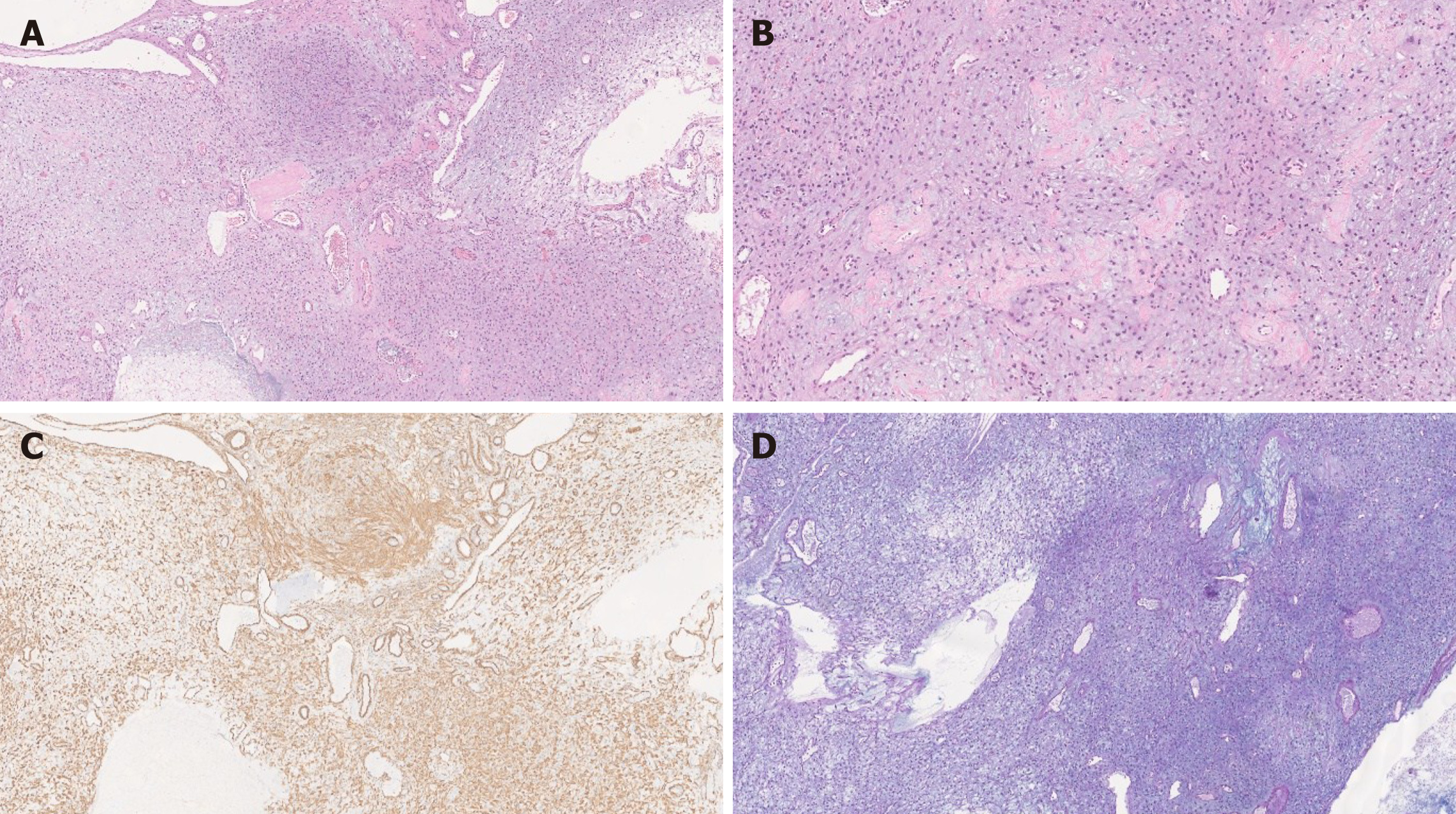
Figure 2 Plexiform fibromyxoma histology.
A: Plexiform fibromyxoma shows characteristic plexiform growth as well as cystic degeneration [hematoxylin and eosin (HE), 50 ×]; B: The tumor is composed of bland spindle to ovoid cells embedded in myxoid stroma, and prominent vasculature (HE, 200 ×); C: The tumor cells are positive for SMA (SMA, 200 ×); D: The myxoid stroma is positive for Alcian Blue (Alcian Blue, 200 ×).
- Citation: Arslan ME, Li H, Fu Z, Jennings TA, Lee H. Plexiform fibromyxoma: Review of rare mesenchymal gastric neoplasm and its differential diagnosis. World J Gastrointest Oncol 2021; 13(5): 409-423
- URL: https://www.wjgnet.com/1948-5204/full/v13/i5/409.htm
- DOI: https://dx.doi.org/10.4251/wjgo.v13.i5.409


