Copyright
©The Author(s) 2021.
World J Gastrointest Oncol. Nov 15, 2021; 13(11): 1680-1695
Published online Nov 15, 2021. doi: 10.4251/wjgo.v13.i11.1680
Published online Nov 15, 2021. doi: 10.4251/wjgo.v13.i11.1680
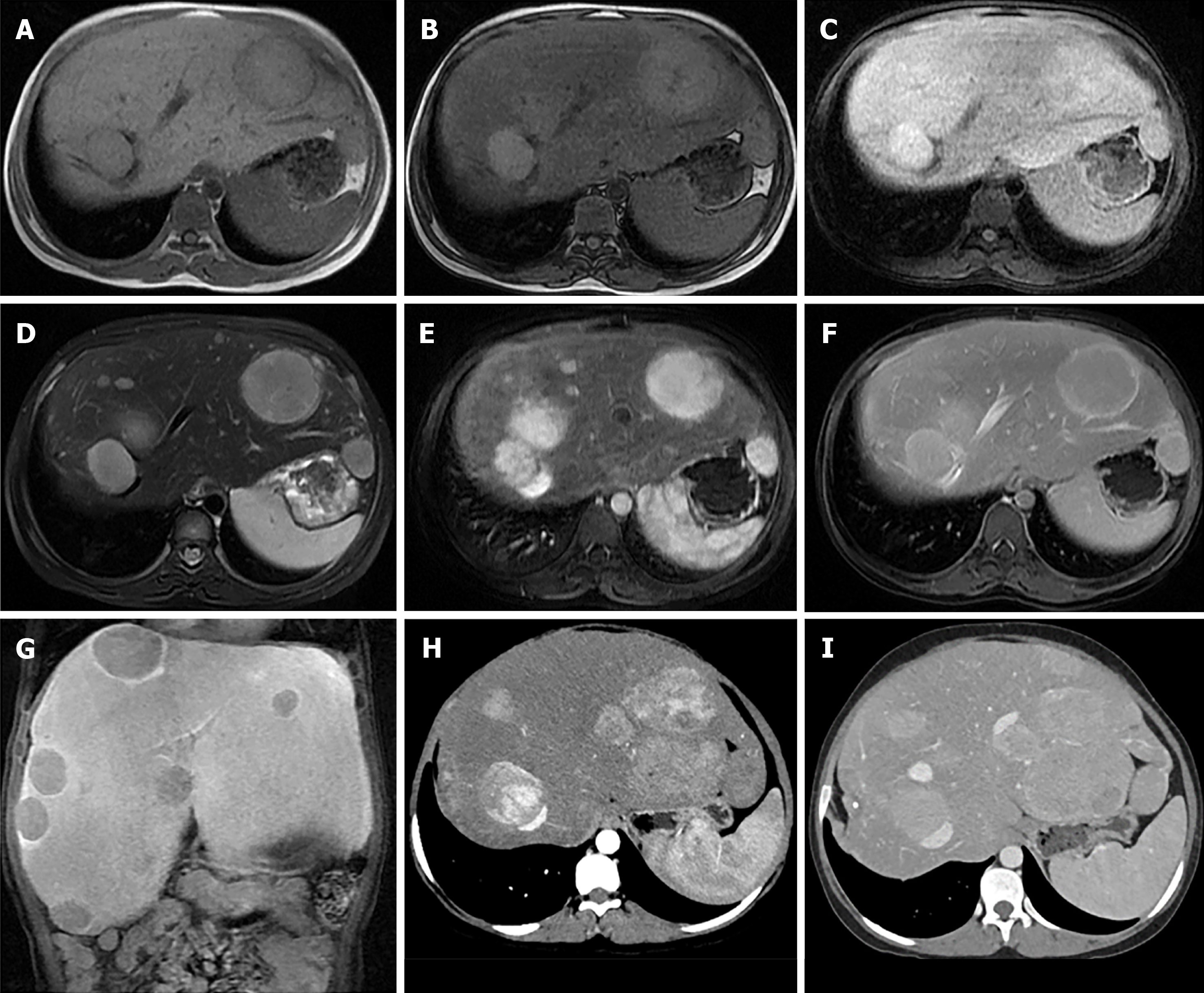
Figure 4 Magnetic resonance images of a 15-year-old girl with underlying GSD type 1 (A-G) and at the 5-yr follow-up, computed tomography was performed (H-I).
One of the identified nodules presented histopathological findings compatible with hepatic adenoma. A and B: Axial dual gradient echo images showing several slightly hyperintense nodules in both hepatic lobes on the background of diffuse hepatic steatosis with dropout in signal intensity of liver parenchyma on contrast-phase images (A) as compared to in-phase images (B); C: Hypointense nodules on the T1-weighted (T1W) image; D: Hyperintense nodules on the T2W image; E-G: Intense arterial hyperenhancement of nodules (E), iso-to mild venous enhancement (F) and hypointense nodules on delayed hepatobiliary phases at 20 min (G) after injection with gadolinium-base hepatocyte specific contrast agent; H: Intense arterial hyperenhancement and increased in size of the nodules; I: Slight hyperenhancement in the venous phase.
- Citation: Sintusek P, Phewplung T, Sanpavat A, Poovorawan Y. Liver tumors in children with chronic liver diseases. World J Gastrointest Oncol 2021; 13(11): 1680-1695
- URL: https://www.wjgnet.com/1948-5204/full/v13/i11/1680.htm
- DOI: https://dx.doi.org/10.4251/wjgo.v13.i11.1680


