Copyright
©The Author(s) 2021.
World J Gastrointest Oncol. Nov 15, 2021; 13(11): 1680-1695
Published online Nov 15, 2021. doi: 10.4251/wjgo.v13.i11.1680
Published online Nov 15, 2021. doi: 10.4251/wjgo.v13.i11.1680
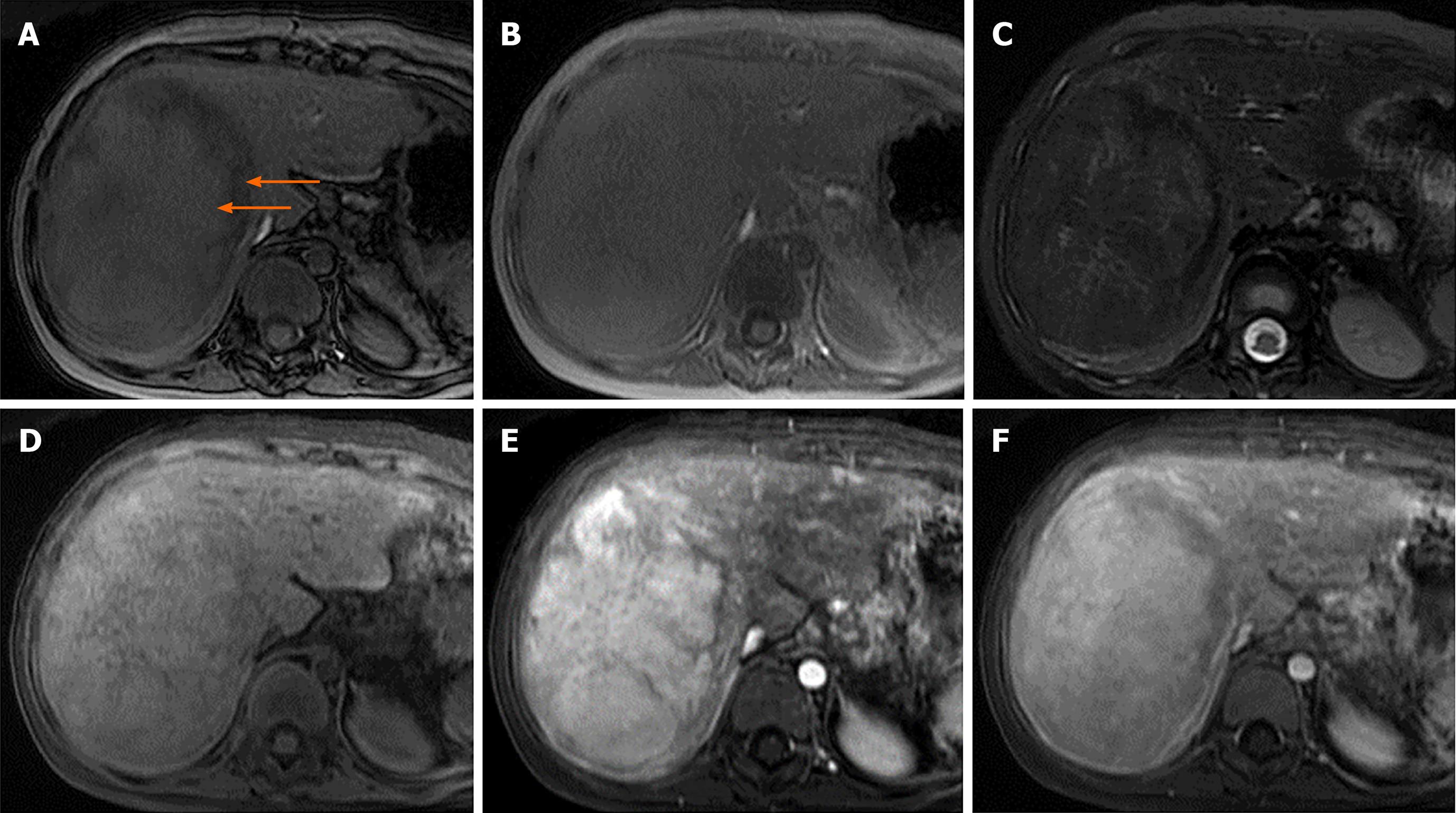
Figure 3 Magnetic resonance images of a 10-year-old girl with extrahepatic hypertension from portal vein thrombosis status post-splenectomy and proximal splenorenal shunt with developing liver mass with pathological tissue diagnosis of hepatic adenoma.
A: Axial dual gradient echo opposed-phase images revealed a heterogeneous drop in signal intensity (arrows); B: Axial dual gradient echo in the in-phase image revealing the heterogeneous microscopic fat in the mass; C: Heterogeneously mild hyperintense mass in T2-weighted (T2W) image; D: Iso-to-slightly hyperintense mass in the T1W image; E: Intense arterial hyperenhancement after gadolinium-based contrast administration; F: Heterogeneous venous enhancement after gadolinium-based contrast administration.
- Citation: Sintusek P, Phewplung T, Sanpavat A, Poovorawan Y. Liver tumors in children with chronic liver diseases. World J Gastrointest Oncol 2021; 13(11): 1680-1695
- URL: https://www.wjgnet.com/1948-5204/full/v13/i11/1680.htm
- DOI: https://dx.doi.org/10.4251/wjgo.v13.i11.1680


