Copyright
©The Author(s) 2020.
World J Gastrointest Oncol. Apr 15, 2020; 12(4): 457-466
Published online Apr 15, 2020. doi: 10.4251/wjgo.v12.i4.457
Published online Apr 15, 2020. doi: 10.4251/wjgo.v12.i4.457
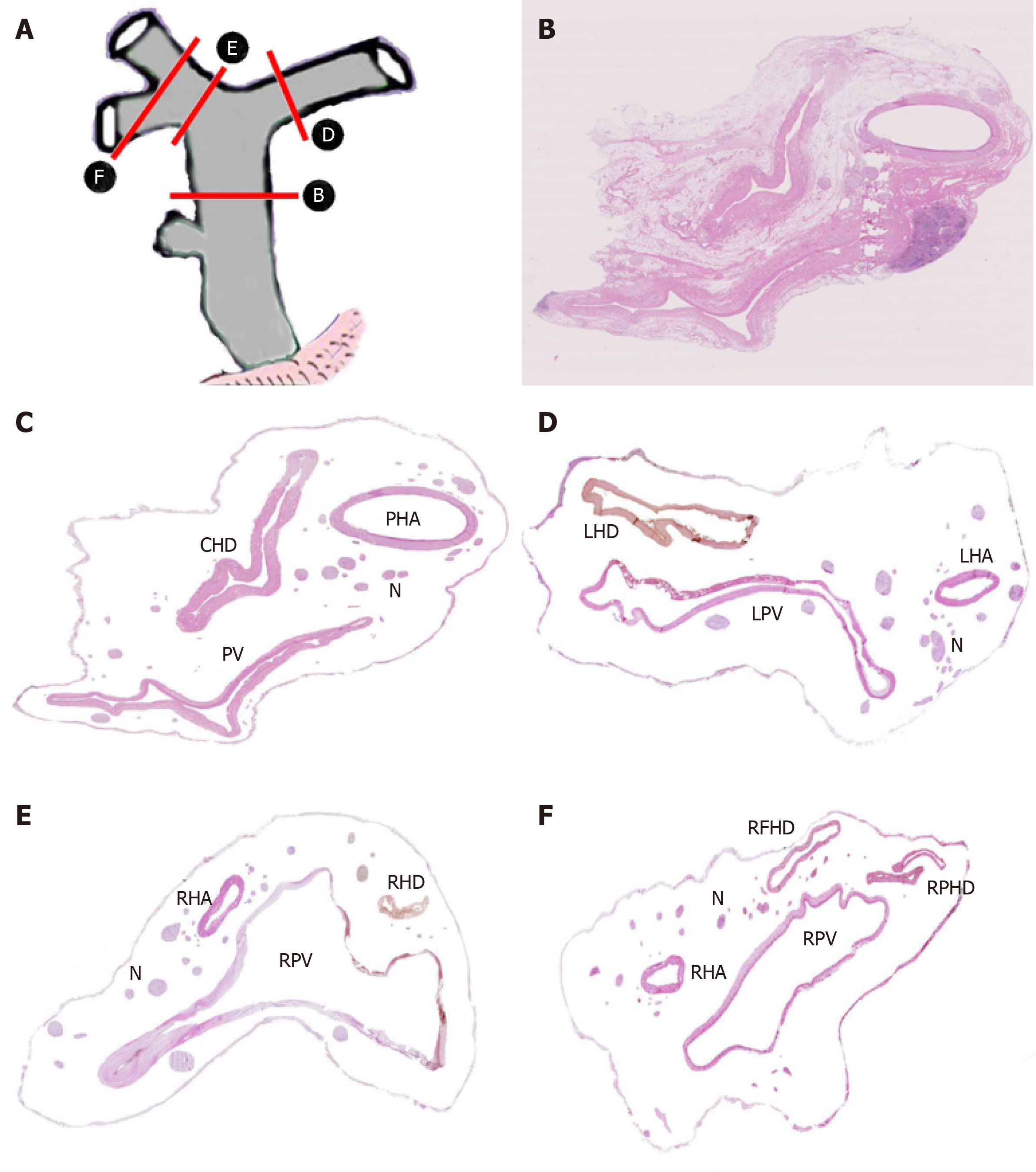
Figure 4 Distribution of nerve plexus around hepatic portal.
A: Delineation of cutting parts; B: Transection of hepatoduodenal ligament including common hepatic duct [Hematoxylin-eosin (HE) staining, original magnification ×200]; C: Figure B without fibrous connective tissue and adipose tissue; D: Transection of the beginning part of Glisson’s sheath, including left hepatic duct (HE staining, original magnification ×200); E: Transection of the beginning part of Glisson’s sheath, including right hepatic duct (HE staining, original magnification ×200); F: Transection of furcation of right hepatic duct (HE staining, original magnification ×200). CHD: Common hepatic duct; PV: Portal vein; PHA: Proper hepatic artery; N: Nerve plexus; LHD: Left hepatic duct; LPV: Left branch of portal vein; LHA: Left hepatic artery; RHD: Right hepatic duct; RPV: Right branch of portal vein; RFID: Right front hepatic duct; RPHD: Right posterior hepatic duct.
- Citation: Li CG, Zhou ZP, Tan XL, Zhao ZM. Perineural invasion of hilar cholangiocarcinoma in Chinese population: One center’s experience. World J Gastrointest Oncol 2020; 12(4): 457-466
- URL: https://www.wjgnet.com/1948-5204/full/v12/i4/457.htm
- DOI: https://dx.doi.org/10.4251/wjgo.v12.i4.457


