Copyright
©The Author(s) 2020.
World J Gastrointest Oncol. Apr 15, 2020; 12(4): 457-466
Published online Apr 15, 2020. doi: 10.4251/wjgo.v12.i4.457
Published online Apr 15, 2020. doi: 10.4251/wjgo.v12.i4.457
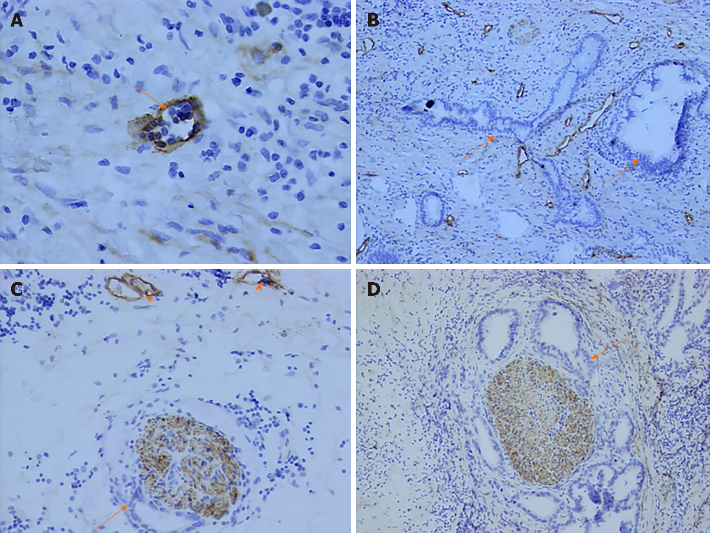
Figure 3 Correlation between perineural invasion and lymphatics.
A: Lymphatic microvessel (arrow) was stained brown (D2-40, original magnification ×400); B: A great of lymph ducts were observed in primary tumor (D2-40, original magnification ×100); C, D: Tumor cells (arrow) invaded nerve fiber and lymph ducts (arrowhead) were stained (D2-40, original magnification ×100).
- Citation: Li CG, Zhou ZP, Tan XL, Zhao ZM. Perineural invasion of hilar cholangiocarcinoma in Chinese population: One center’s experience. World J Gastrointest Oncol 2020; 12(4): 457-466
- URL: https://www.wjgnet.com/1948-5204/full/v12/i4/457.htm
- DOI: https://dx.doi.org/10.4251/wjgo.v12.i4.457


