Copyright
©The Author(s) 2020.
World J Gastrointest Oncol. Mar 15, 2020; 12(3): 248-266
Published online Mar 15, 2020. doi: 10.4251/wjgo.v12.i3.248
Published online Mar 15, 2020. doi: 10.4251/wjgo.v12.i3.248
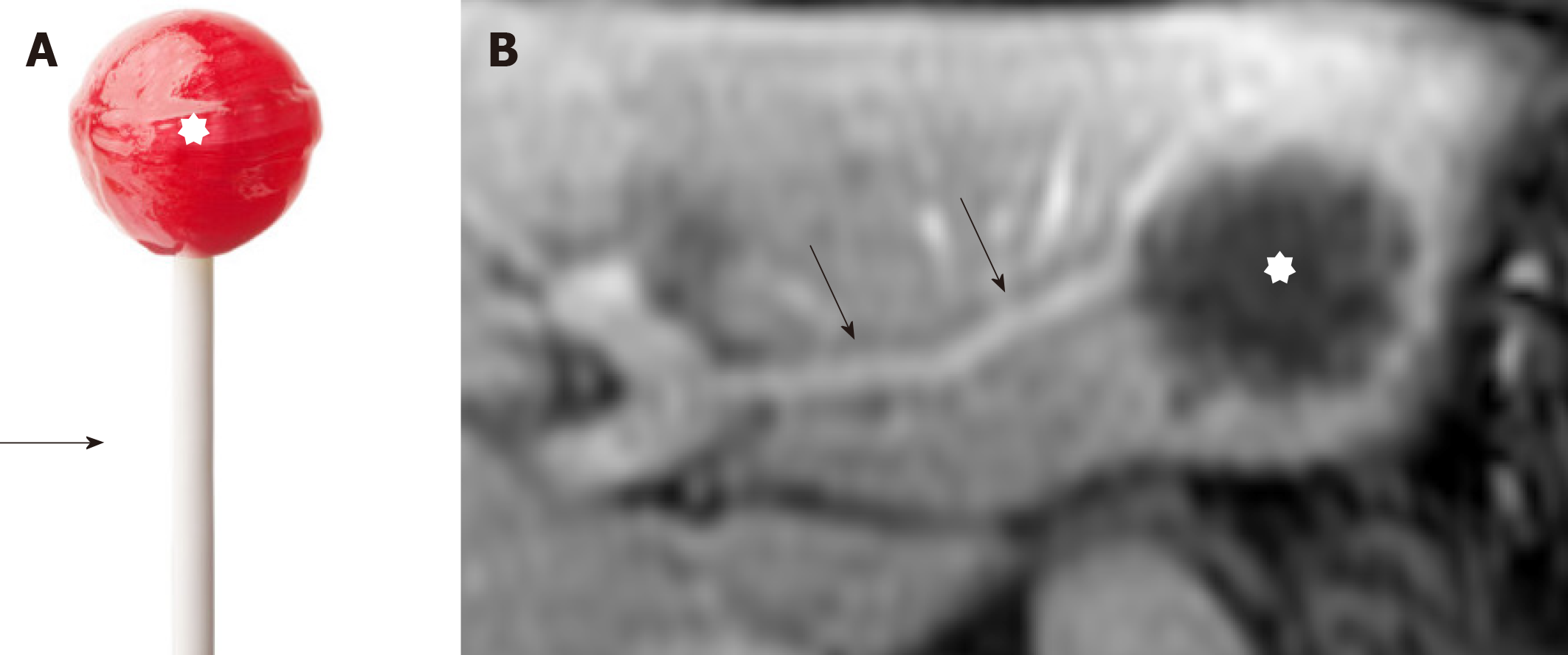
Figure 3 Hepatic hemangioepithelioma imaging feature.
A: A lollipop; B: Axial post contrast T1 Weighted Images images. Hepatic hemangioepithelioma nodule (star) with portal veins entering and terminating in the periphery of the lesion (arrow). This configuration resembles a lollipop. The ‘‘Lollipop sign’’ is a combination of two structures: the well-defined tumor mass on enhanced images (the candy in the lollipop) and the adjacent occluded vein (the stick), because hepatic hemangioepithelioma has the tendency to spread within the portal and hepatic vein branches. The vein should terminate smoothly at the edge or just within the rim of the lesion; vessels that traverse the entire lesion or are displaced and collateral veins cannot be included in the sign.
- Citation: Virarkar M, Saleh M, Diab R, Taggart M, Bhargava P, Bhosale P. Hepatic Hemangioendothelioma: An update. World J Gastrointest Oncol 2020; 12(3): 248-266
- URL: https://www.wjgnet.com/1948-5204/full/v12/i3/248.htm
- DOI: https://dx.doi.org/10.4251/wjgo.v12.i3.248


