Copyright
©The Author(s) 2020.
World J Gastrointest Oncol. Mar 15, 2020; 12(3): 248-266
Published online Mar 15, 2020. doi: 10.4251/wjgo.v12.i3.248
Published online Mar 15, 2020. doi: 10.4251/wjgo.v12.i3.248
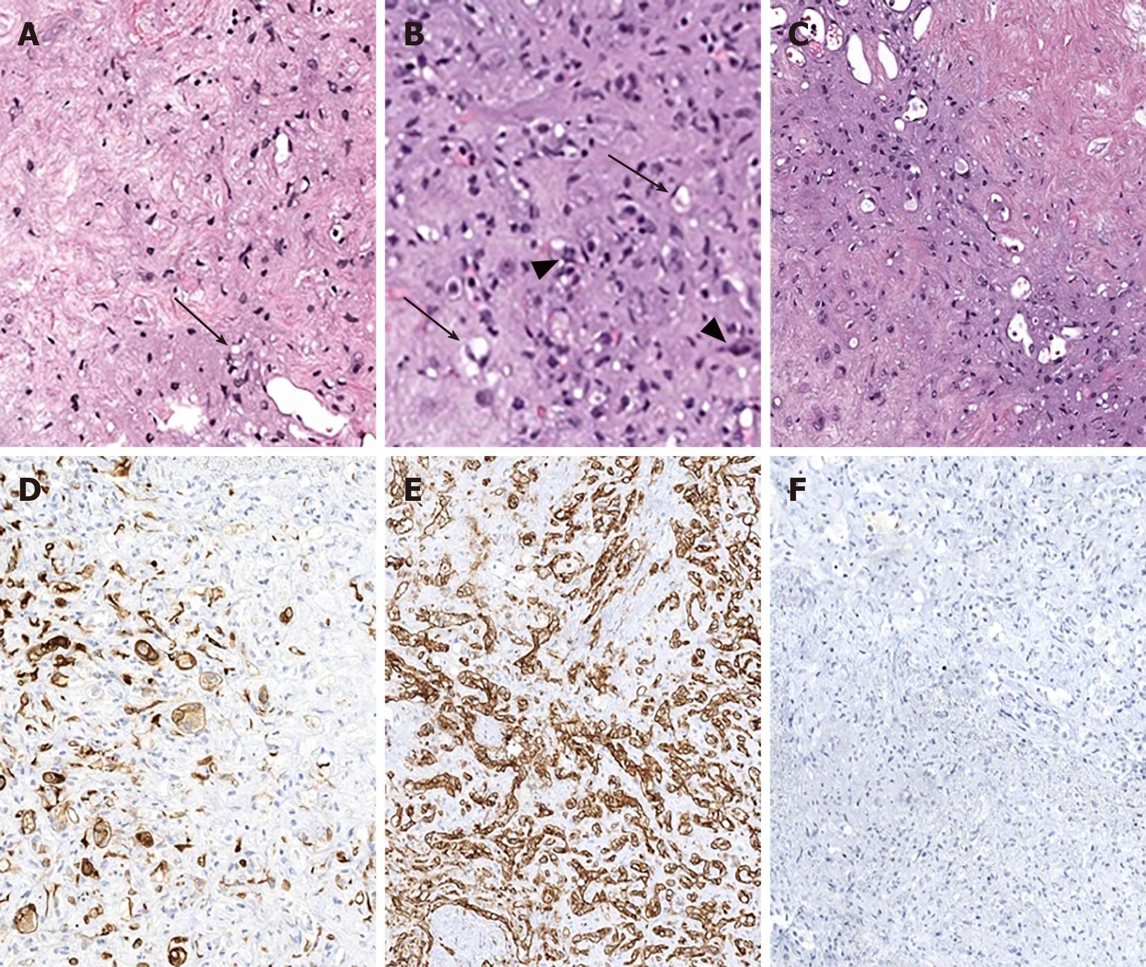
Figure 1 Epithelioid hemangioendothelioma microscopic features.
There is variable cellularity, ranging from stromal-rich areas, mimicking cartilage to highly cellular regions. A: The tumor consists mostly of stellate cells with only rare epithelioid cells containing intracytoplasmic lumen/vacuoles (arrows); B: In more cellular areas, the intracellular lumens (arrows) are more prominent. In addition, the tumor infiltrates sinusoids forming tufted foci (arrowheads); C: Transitional areas between fibrous and moderately cellular areas. To confirm and differentiate epithelioid hemangioendothelioma from tumors with similar histologic features (most commonly cholangiocarcinoma, hepatocellular carcinoma and metastatic signet ring cell carcinoma), immunohistochemical stains are needed; D: Patchy staining for cytokeratin 7, a stain commonly expressed in adenocarcinomas of the upper gastrointestinal tract and pancreaticobiliary tree; E: Diffusely positive for the endothelial marker cluster of differentiation-31 (CD31); F: Negative for the hepatocellular marker, Hepatocyte Paraffin 1 (HepPar1). CD31: Cluster of differentiation-31; HepPar1: Hepatocyte Paraffin 1.
- Citation: Virarkar M, Saleh M, Diab R, Taggart M, Bhargava P, Bhosale P. Hepatic Hemangioendothelioma: An update. World J Gastrointest Oncol 2020; 12(3): 248-266
- URL: https://www.wjgnet.com/1948-5204/full/v12/i3/248.htm
- DOI: https://dx.doi.org/10.4251/wjgo.v12.i3.248


