Copyright
©The Author(s) 2020.
World J Gastrointest Oncol. Dec 15, 2020; 12(12): 1407-1415
Published online Dec 15, 2020. doi: 10.4251/wjgo.v12.i12.1407
Published online Dec 15, 2020. doi: 10.4251/wjgo.v12.i12.1407
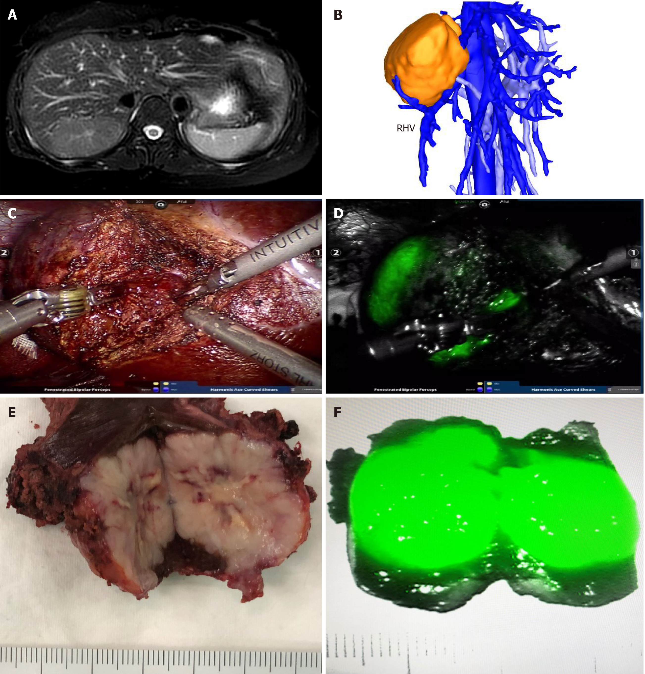
Figure 1 Robotic resection of liver focal nodal hyperplasia guided by indocyanine green fluorescence imaging.
A: Magnetic resonance imaging showed focal nodal hyperplasia (FNH) located in segment VII; B: Three-dimensional reconstruction of computed tomography showed the relationship of FNH and the right hepatic vein; C and D: Robotic resection of liver FNH guided by indocyanine green (ICG) fluorescence imaging; E and F: The resected specimen and the mode of ICG imaging in FNH. RHV: Right hepatic vein.
- Citation: Li CG, Zhou ZP, Tan XL, Wang ZZ, Liu Q, Zhao ZM. Robotic resection of liver focal nodal hyperplasia guided by indocyanine green fluorescence imaging: A preliminary analysis of 23 cases. World J Gastrointest Oncol 2020; 12(12): 1407-1415
- URL: https://www.wjgnet.com/1948-5204/full/v12/i12/1407.htm
- DOI: https://dx.doi.org/10.4251/wjgo.v12.i12.1407


