Copyright
©The Author(s) 2020.
World J Gastrointest Oncol. Nov 15, 2020; 12(11): 1336-1345
Published online Nov 15, 2020. doi: 10.4251/wjgo.v12.i11.1336
Published online Nov 15, 2020. doi: 10.4251/wjgo.v12.i11.1336
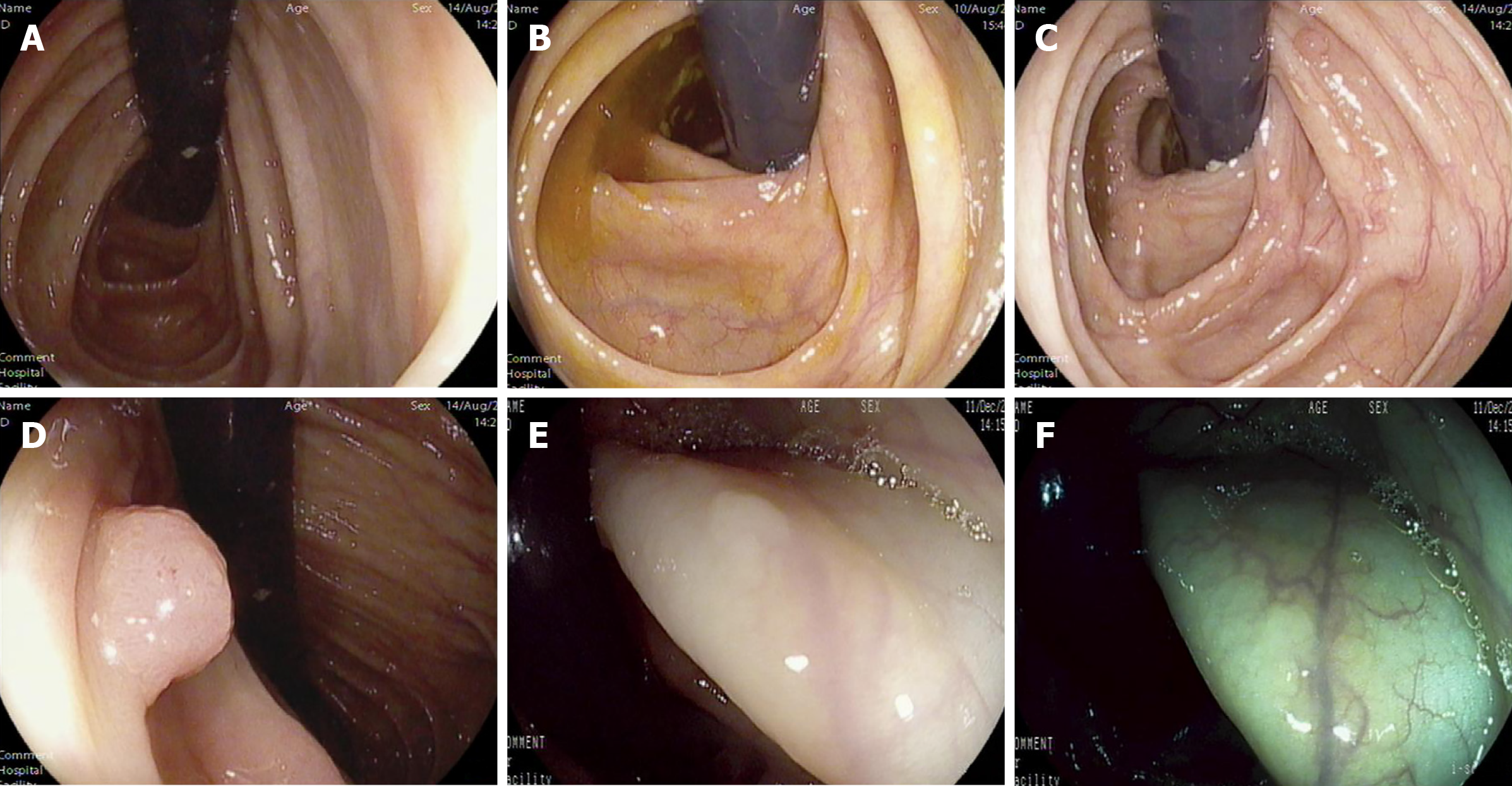
Figure 3 Detected lesions.
A and B: The colonoscopy was successfully retroflexed in the ascending colon and the hepatic flexure, respectively; C and D: The same polyp hidden in the proximal side of the haustral folds in the distant view and the tight view, respectively; E and F: A flat lesion located on the haustral folds with white-light mode and i-SCAN mode, respectively.
- Citation: Li WK, Wang Y, Wang YD, Liu KL, Guo CM, Su H, Liu H, Wu J. Diagnostic value of novel retroflexion colonoscopy in the right colon: A randomized controlled trial. World J Gastrointest Oncol 2020; 12(11): 1336-1345
- URL: https://www.wjgnet.com/1948-5204/full/v12/i11/1336.htm
- DOI: https://dx.doi.org/10.4251/wjgo.v12.i11.1336


