Copyright
©The Author(s) 2020.
World J Gastrointest Oncol. Nov 15, 2020; 12(11): 1216-1236
Published online Nov 15, 2020. doi: 10.4251/wjgo.v12.i11.1216
Published online Nov 15, 2020. doi: 10.4251/wjgo.v12.i11.1216
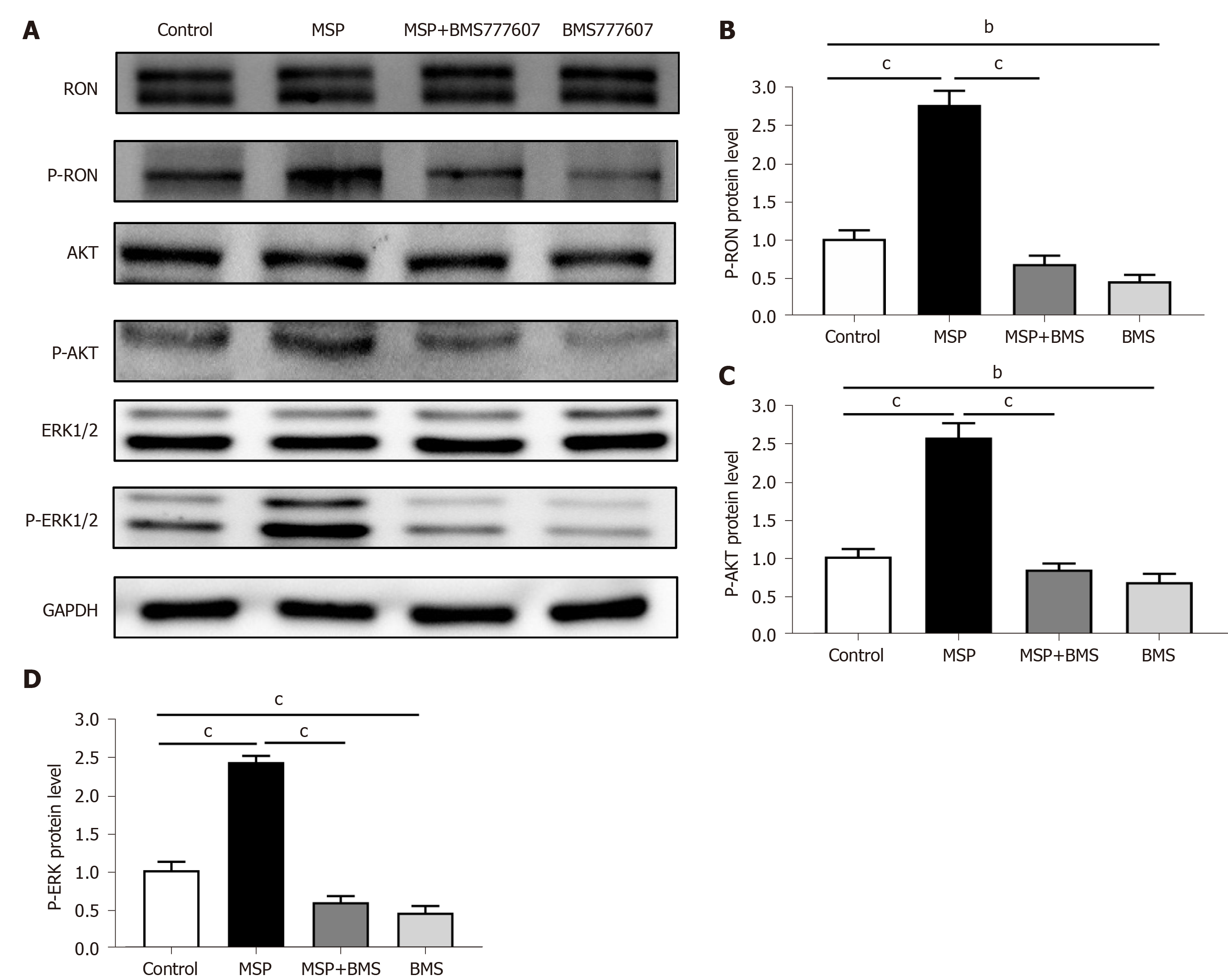
Figure 6 Expression of RON and PD-L1, and activation of signaling pathways in HT29 cells.
A: HT29 cells were treated with 2 nmol/L MSP, 2 nmol/L MSP + 2 μmol/L BMS-777607, or 2 μmol/L BMS-777607 for 1 h. The proteins analyzed include RON, phosphorylated RON, AKT, ERK1/2, phosphorylated AKT, and phosphorylated-ERK1/2. GAPDH was used as a loading control; B-D: The expression of p-RON, p-AKT, and p-ERK1/2 was detected by Western blot and quantified. aP < 0.05, bP < 0.01, cP < 0.001.
- Citation: Liu YZ, Han DT, Shi DR, Hong B, Qian Y, Wu ZG, Yao SH, Tang TM, Wang MH, Xu XM, Yao HP. Pathological significance of abnormal recepteur d’origine nantais and programmed death ligand 1 expression in colorectal cancer. World J Gastrointest Oncol 2020; 12(11): 1216-1236
- URL: https://www.wjgnet.com/1948-5204/full/v12/i11/1216.htm
- DOI: https://dx.doi.org/10.4251/wjgo.v12.i11.1216


