Copyright
©The Author(s) 2019.
World J Gastrointest Oncol. Sep 15, 2019; 11(9): 761-767
Published online Sep 15, 2019. doi: 10.4251/wjgo.v11.i9.761
Published online Sep 15, 2019. doi: 10.4251/wjgo.v11.i9.761
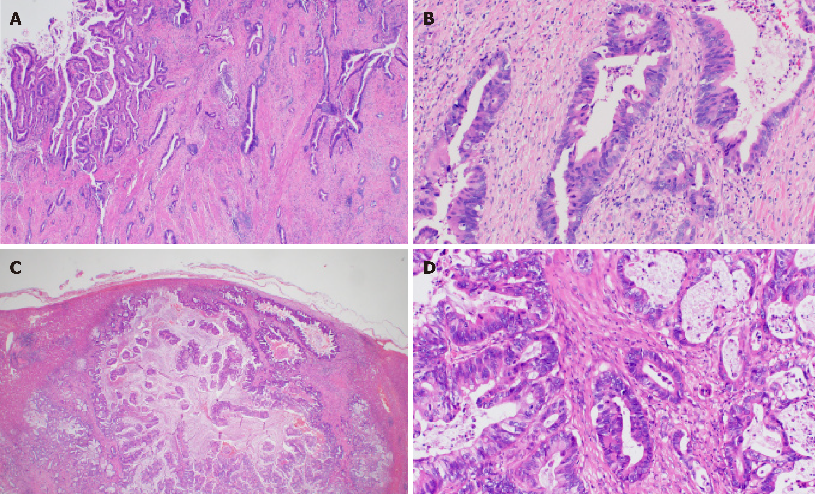
Figure 2 Tumor histology.
A: Low-power microscopic view of the gallbladder cancer. The tumor cells form tubules of variable sizes. The tumor infiltrates deeply into the gallbladder wall; B: High-power microscopic view of the gallbladder cancer. Atypical columnar cells with enlarged nuclei grow in tubular structures. Stromal fibrosis and inflammation are also observed; C: Low-power microscopic view of the hepatic lesion. Adenocarcinoma with vaguely nodular contour involves the liver parenchyma; D: High-power microscopic view of the hepatic lesion. Glandular structure is predominant. The tumor cells have enlarged nuclei with coarse chromatin. Multiple mitoses are observed.
- Citation: Inagaki C, Maeda D, Kimura A, Otsuru T, Iwagami Y, Nishida N, Sakai D, Shitotsuki R, Yachida S, Doki Y, Satoh T. Gallbladder cancer harboring ERBB2 mutation on the primary and metastatic site: A case report. World J Gastrointest Oncol 2019; 11(9): 761-767
- URL: https://www.wjgnet.com/1948-5204/full/v11/i9/761.htm
- DOI: https://dx.doi.org/10.4251/wjgo.v11.i9.761


